Quantification of Initial Right Ventricular Dimensions by Computed Tomography in Infants with Congenital Heart Disease and a Hypoplastic Right Ventricle
- Affiliations
-
- 1Department of Radiology and Research Institute of Radiology, University of Ulsan College of Medicine, Asan Medical Center, Seoul, Korea. ghw68@hanmail.net
- KMID: 2471637
- DOI: http://doi.org/10.3348/kjr.2019.0662
Abstract
OBJECTIVE
To demonstrate the feasibility of using cardiothoracic CT for quantification of the initial right ventricle (RV) dimensions in infants with congenital heart disease (CHD) and a hypoplastic RV and to compare these measurements with those obtained in a control group with CHD without a hypoplastic RV.
MATERIALS AND METHODS
Initial RV dimensions, including RV volumes, RV/left ventricle (LV) volume ratios, atrioventricular valve annulus diameter ratios, and RV/LV length ratios based on CT data, were collected from 57 infants with CHD and a hypoplastic RV (hypoplastic RV group; age range, 1 day to 6 months) and 33 infants with tetralogy of Fallot (control group; age range, 1 day to 6 months) and compared between the 2 groups. The type of final surgery was also evaluated in the hypoplastic RV group over a follow-up period of 3-8 years.
RESULTS
The RV and LV volumes and lengths were successfully quantified in all 90 patients. The tricuspid valve annulus diameter could not be measured in cases showing muscular tricuspid atresia and double-inlet LV. The initial RV dimensions quantified by CT were significantly lower for the hypoplastic RV group than for the control group (p < 0.001). The types of final surgery performed in the hypoplastic RV group were univentricular repair in 46 patients, biventricular repair in 4 patients, or an indeterminate surgery in 7 patients.
CONCLUSION
Initial RV dimensions in infants with CHD and a hypoplastic RV can be quantified by CT and are substantially smaller than those in infants with tetralogy of Fallot.
Keyword
MeSH Terms
Figure
-
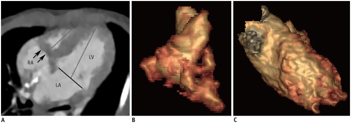
Fig. 1 Initial cardiothoracic CT in 8-day-old female newborn with muscular tricuspid atresia.A. Four-chamber image shows muscular atresia (arrows) of tricuspid valve and hypoplastic RV. Mitral valve annulus diameter (black line) and length of RV and LV (gray lines) were measurable. Tricuspid valve annulus diameter was not measurable. As result, atrioventricular valve annulus diameter ratio could not be calculated. B. RV ESVi quantified with CT ventricular volumetry was 4.7 mL/m2. C. LV ESVi quantified with CT ventricular volumetry was 30.0 mL/m2.CT = computed tomography, ESVi = indexed end-systolic volume, LA = left atrium, LV = left ventricle, RA = right atrium, RV = right ventricle
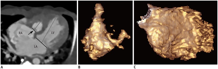
Fig. 2 Initial cardiothoracic CT in 4-day-old female newborn with tricuspid atresia with imperforate valve.A. Four-chamber image shows imperforated tricuspid valve (arrow) and hypoplastic RV. In contrast to muscular type of tricuspid atresia, annulus diameters (black lines) of tricuspid and mitral valves as well as length of RV and LV (gray lines) were measurable. B. RV ESVi quantified with CT ventricular volumetry was 6.2 mL/m2. C. LV ESVi quantified with CT ventricular volumetry was 39.0 mL/m2.
Reference
-
1. Muster AJ, Zales VR, Ilbawi MN, Backer CL, Duffy CE, Mavroudis C. Biventricular repair of hypoplastic right ventricle assisted by pulsatile bidirectional cavopulmonary anastomosis. J Thorac Cardiovasc Surg. 1993; 105:112–119. PMID: 8419691.
Article2. Gentles TL, Keane JF, Jonas RA, Marx GE, Mayer JE Jr. Surgical alternatives to the Fontan procedure incorporating a hypoplastic right ventricle. Circulation. 1994; 90(5 Pt 2):II1–II6.3. De Oliveira NC, Sittiwangkul R, McCrindle BW, Dipchand A, Yun TJ, Coles JG, et al. Biventricular repair in children with atrioventricular septal defects and a small right ventricle: anatomic and surgical considerations. J Thorac Cardiovasc Surg. 2005; 130:250–257. PMID: 16077383.4. Yoshimura N, Yamaguchi M. Surgical strategy for pulmonary atresia with intact ventricular septum: initial management and definitive surgery. Gen Thorac Cardiovasc Surg. 2009; 57:338–346. PMID: 19597923.
Article5. Awori MN, Mehta NP, Mitema FO, Kebba N. Optimal Z-score use in surgical decision-making in pulmonary atresia with intact ventricular septum. World J Pediatr Congenit Heart Surg. 2017; 8:385–388. PMID: 28520535.
Article6. Chen RHS, Chau AKT, Chow PC, Yung TC, Cheung YF, Lun KS. Achieving biventricular circulation in patients with moderate hypoplastic right ventricle in pulmonary atresia intact ventricular septum after transcatheter pulmonary valve perforation. Congenit Heart Dis. 2018; 13:884–891. PMID: 30238621.
Article7. Goo HW, Park SH. Semiautomatic three-dimensional CT ventricular volumetry in patients with congenital heart disease: agreement between two methods with different user interaction. Int J Cardiovasc Imaging. 2015; 31 Suppl 2:223–232. PMID: 26319216.
Article8. Kim HJ, Goo HW, Park SH, Yun TJ. Left ventricle volume measured by cardiac CT in an infant with a small left ventricle: a new and accurate method in determining uni- or biventricular repair. Pediatr Radiol. 2013; 43:243–246. PMID: 22875206.
Article9. Goo HW. Serial changes in anatomy and ventricular function on dual-source cardiac computed tomography after the Norwood procedure for hypoplastic left heart syndrome. Pediatr Radiol. 2017; 47:1776–1786. PMID: 28879411.
Article10. Goo HW, Park SH. Computed tomography-based ventricular volumes and morphometric parameters for deciding the treatment strategy in children with a hypoplastic left ventricle: preliminary results. Korean J Radiol. 2018; 19:1042–1052. PMID: 30386136.
Article11. Goo HW, Suh DS. Tube current reduction in pediatric non-ECG-gated heart CT by combined tube current modulation. Pediatr Radiol. 2006; 36:344–351. PMID: 16501970.
Article12. Goo HW. State-of-the-art CT imaging techniques for congenital heart disease. Korean J Radiol. 2010; 11:4–18. PMID: 20046490.
Article13. Goo HW. Individualized volume CT dose index determined by cross-sectional area and mean density of the body to achieve uniform image noise of contrast-enhanced pediatric chest CT obtained at variable kV levels and with combined tube current modulation. Pediatr Radiol. 2011; 41:839–847. PMID: 21656275.
Article14. Goo HW. Is it better to enter a volume CT dose index value before or after scan range adjustment for radiation dose optimization of pediatric cardiothoracic CT with tube current modulation? Korean J Radiol. 2018; 19:692–703. PMID: 29962875.
Article15. Hong SH, Goo HW, Maeda E, Choo KS, Tsai IC;. User-friendly vendor-specific guideline for pediatric cardiothoracic computed tomography provided by the Asian Society of Cardiovascular Imaging Congenital Heart Disease Study Group: part 1. Imaging techniques. Korean J Radiol. 2019; 20:190–204. PMID: 30672159.
Article16. Tricarico F, Hlavacek AM, Schoepf UJ, Ebersberger U, Nance JW Jr, Vliegenthart R, et al. Cardiovascular CT angiography in neonates and children: image quality and potential for radiation dose reduction with iterative image reconstruction techniques. Eur Radiol. 2013; 23:1306–1315. PMID: 23207869.
Article17. Goo HW. Combined prospectively electrocardiography- and respiratory-triggered sequential cardiac computed tomography in free-breathing children: success rate and image quality. Pediatr Radiol. 2018; 48:923–931. PMID: 29589058.
Article18. Goo HW. Semiautomatic three-dimensional threshold-based cardiac computed tomography ventricular volumetry in repaired tetralogy of Fallot: comparison with cardiac magnetic resonance imaging. Korean J Radiol. 2019; 20:102–113. PMID: 30627026.
Article19. Shiota T. 3D echocardiography: evaluation of the right ventricle. Curr Opin Cardiol. 2009; 24:410–414. PMID: 19550311.
Article20. Oshima Y, Maruo A, Hasegawa T, Matsuhisa H. Repair of unbalanced atrioventricular septal defect with small right ventricle. Asian Cardiovasc Thorac Ann. 2016; 24:78–80. PMID: 25061220.
Article21. Huang SC, Ishino K, Kasahara S, Yoshizumi K, Kotani Y, Sano S. The potential of disproportionate growth of tricuspid valve after decompression of the right ventricle in patients with pulmonary atresia and intact ventricular septa. J Thorac Cardiovasc Surg. 2009; 138:1160–1166. PMID: 19837219.
Article
- Full Text Links
-
- Actions
-
Cited
- CITED
-
- Close
- Share
-
- Similar articles
-
- 2 Cases of Hypoplastic Left Heart Syndrome
- Noncompaction of Ventricular Myocardium Involving the Right Ventricle
- Three-Dimensional Endo-Cardiovascular Volume-Rendered Cine Computed Tomography of Isolated Left Ventricular Apical Hypoplasia: A Case Report and Literature Review
- Pattern Analysis of Left Ventricular Remodeling Using Cardiac Computed Tomography in Children with Congenital Heart Disease: Preliminary Results
- Functional Assessment for Congenital Heart Disease
