Full mouth rehabilitation of a severely worn dentition using intraoral scanner and the CAD/CAM double scanning technique
- Affiliations
-
- 1Department of Prosthodontics, School of Dentistry, Seoul National University, Seoul, Republic of Korea. drhiy226@snu.ac.kr
- KMID: 2469923
- DOI: http://doi.org/10.4047/jkap.2020.58.1.67
Abstract
- With the evolution of the computer-aided design/computer-aided manufacturing (CAD/CAM) technology, the intraoral scanners are playing an increasingly important role, as they are the first step towards a completely digital workflow. The CAD/CAM double scanning technique has been used to transfer the information from provisional restorations to definitive restorations. In this case, a 67-year-old male with esthetically compromised anterior teeth, generalized severe attrition of teeth, and reduced vertical dimension was treated with full mouth rehabilitation including a re-establishment of the lost vertical dimension of occlusion assisted by the crown lengthening procedure. The provisional restorations were fabricated using an intraoral scanner and the CAD/CAM double scanning technique. After the period of adaption, the definitive monolithic zirconia restorations were delivered. The CAD/CAM double scanning technique successfully transferred the occlusal and morphological characteristics, obtained from the provisional restorations, to the definitive restorations.
Keyword
MeSH Terms
Figure
-
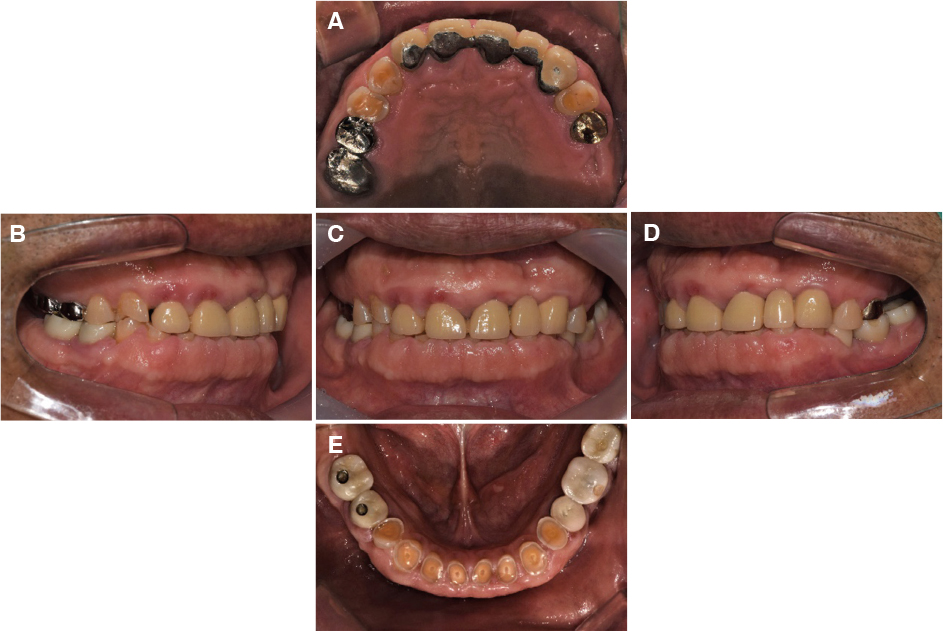
Fig. 1 Intraoral photograph before treatment. (A) Maxillary occlusal, (B) Right, (C) Frontal, (D) Left, (E) Mandibular occlusal views.
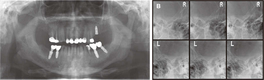
Fig. 2 Initial radiographs of the patient. (A) Panoramic radiograph, (B) Transcranial projection of both TMJ.

Fig. 3 Occlusal splint with re-establishment of the vertical dimension.
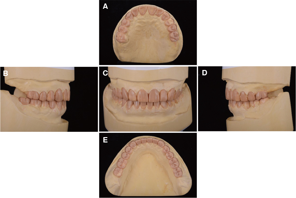
Fig. 4 Diagnostic wax-up models. (A) Maxillary occlusal, (B) Right, (C) Frontal, (D) Left, (E) Mandibular occlusal views.
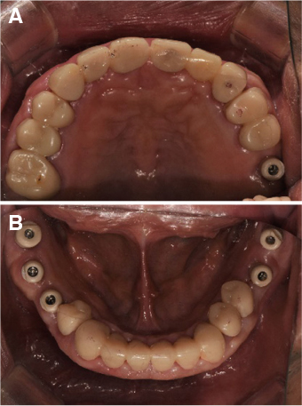
Fig. 5 Intraoral scanbody. (A) Maxillary, (B) Mandibular.
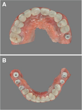
Fig. 6 Intraoral scan. (A) Maxillary, (B) Mandibular.
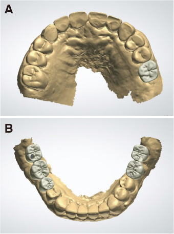
Fig. 7 CAD design. (A) Maxillary, (B) Mandibular.
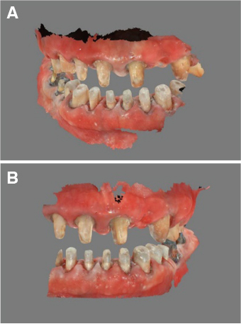
Fig. 8 Two-thirds of the arch with the prepared teeth and titanium abutments were scanned. (A) Right, (B) Left.

Fig. 9 Two-thirds of the arch with the provisional restorations were scanned. (A) Right, (B) Left.

Fig. 10 Superimposition of the prepared teeth, titanium abutments, and provisional restorations. (A) Right, (B) Left.

Fig. 11 Second provisional restorations. (A) Maxillary occlusal, (B) Right, (C) Frontal, (D) Left, (E) Mandibular occlusal views.

Fig. 12 Final impression of the anterior teeth abutments. (A) Maxillary, (B) Mandibular.
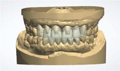
Fig. 13 Superimposition of second provisional restorations and the working casts (anterior teeth).
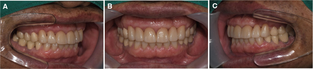
Fig. 14 Definitive restorations of the anterior teeth. (A) Right, (B) Frontal, (C) Left.
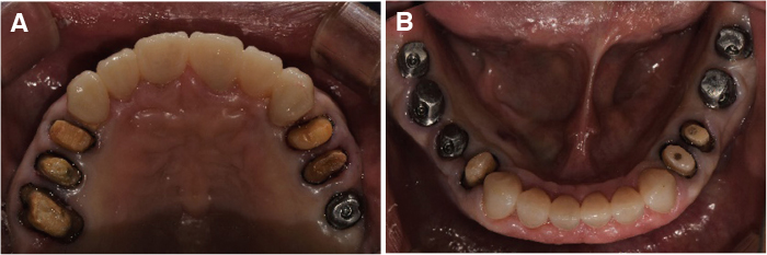
Fig. 15 Final impression of the posterior teeth abutments. (A) Maxillary, (B) Mandibular.

Fig. 16 Superimposition of second provisional restorations and the working casts (posterior teeth).
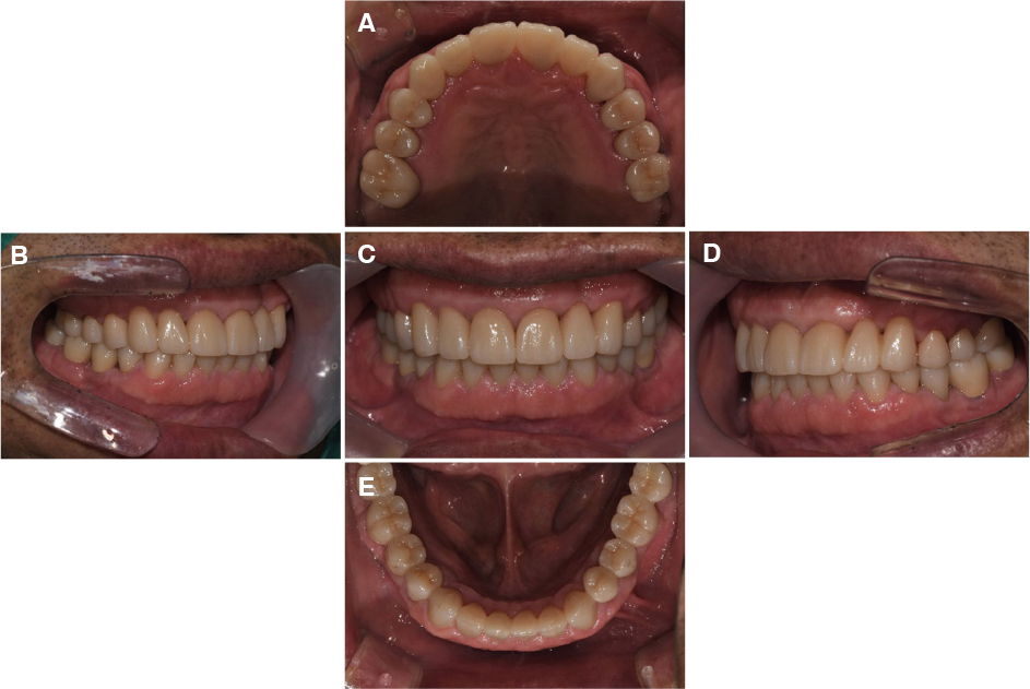
Fig. 17 Definitive restorations. (A) Maxillary occlusal, (B) Right, (C) Frontal, (D) Left, (E) Mandibular occlusal views

Fig. 18 Panoramic radiograph after treatment.
Reference
-
1. Turner KA, Missirlian DM. Restoration of the extremely worn dentition. J Prosthet Dent. 1984; 52:467–474.
Article2. Gopi Chander N, Venkat R. An appraisal on increasing the occlusal vertical dimension in full occlusal rehabilitation and its outcome. J Indian Prosthodont Soc. 2011; 11:77–81.
Article3. Song MY, Park JM, Park EJ. Full mouth rehabilitation of the patient with severely worn dentition: a case report. J Adv Prosthodont. 2010; 2:106–110.
Article4. Miyazaki T, Hotta Y, Kunii J, Kuriyama S, Tamaki Y. A review of dental CAD/CAM: current status and future perspectives from 20 years of experience. Dent Mater J. 2009; 28:44–56.
Article5. Nedelcu R, Olsson P, Nyström I, Rydén J, Thor A. Accuracy and precision of 3 intraoral scanners and accuracy of conventional impressions: A novel in vivo analysis method. J Dent. 2018; 69:110–118.
Article6. Amin S, Weber HP, Finkelman M, El Rafie K, Kudara Y, Papaspyridakos P. Digital vs. conventional full-arch implant impressions: a comparative study. Clin Oral Implants Res. 2017; 28:1360–1367.
Article7. Ender A, Attin T, Mehl A. In vivo precision of conventional and digital methods of obtaining complete-arch dental impressions. J Prosthet Dent. 2016; 115:313–320.
Article8. Ueda K, Güth JF, Erdelt K, Stimmelmayr M, Kappert H, Beuer F. Light transmittance by a multi-coloured zirconia material. Dent Mater J. 2015; 34:310–314.
Article9. Baldissara P, Wandscher VF, Marchionatti AME, Parisi C, Monaco C, Ciocca L. Translucency of IPS e.max and cubic zirconia monolithic crowns. J Prosthet Dent. 2018; 120:269–275.
Article10. Joo HS, Park SW, Yun KD, Lim HP. Complete-mouth rehabilitation using a 3D printing technique and the CAD/CAM double scanning method: A clinical report. J Prosthet Dent. 2016; 116:3–7.
Article11. Kim JY, Park SW, Lim HP, Yun KD, Yang H. Rehabilitation in a patient with limited restorable space using double scanning technique: A case report. J Korean Acad Prosthodont. 2017; 55:205–211.
Article12. Song JW, Leesungbok R, Park SJ, Chang SH, Ahn SJ, Lee SW. Analysis of crown size and morphology, and gingival shape in the maxillary anterior dentition in Korean young adults. J Adv Prosthodont. 2017; 9:315–320.
Article13. Patzelt SB, Emmanouilidi A, Stampf S, Strub JR, Att W. Accuracy of full-arch scans using intraoral scanners. Clin Oral Investig. 2014; 18:1687–1694.
Article14. Su TS, Sun J. Comparison of repeatability between intraoral digital scanner and extraoral digital scanner: An in-vitro study. J Prosthodont Res. 2015; 59:236–242.
Article
- Full Text Links
-
- Actions
-
Cited
- CITED
-
- Close
- Share
-
- Similar articles
-
- Full mouth rehabilitation in a patient with reduced vertical dimension due to numerous tooth loss and excessie worn dentition: A case report
- Computer-aided design and manufacturing-based full mouth rehabilitation for a patient with excessive attrition and restricted vertical dimension: A case report
- Full mouth rehabilitation using zirconia crown in severe worn dentition: a case report
- Application of various digital technique on full mouth rehabilitation: A case report
- Integrating 3D facial scanning in a digital workflow to CAD/CAM design and fabricate complete dentures for immediate total mouth rehabilitation
