Full mouth rehabilitation in a patient with reduced vertical dimension due to numerous tooth loss and excessie worn dentition: A case report
- Affiliations
-
- 1Department of Prosthodontics, School of Dentistry, Seoul National University, Seoul, Republic of Korea. silk1@snu.ac.kr
- KMID: 2461154
- DOI: http://doi.org/10.4047/jkap.2019.57.4.456
Abstract
- As digital dentistry technology is being developed, it is being used in various ways. This case covers how digital dentistry technology is being applied on the treatment of patients with loss of vertical dimension due to worn dentition and multiple loss of teeth. The loss of vertical dimension was carefully assessed and recovered, and implants were placed with surgical guides, designed considering the final restoration. The movement of the mandibular was measured with the electronic instrument for recording mandibular movement. Wax-up process was done with Naturgemäße Aufwachs-Technik (N.A.T.) and Natural functional reconstruction (N.F.R.). It was scanned, and the provisional restoration was fabricated using Computer-Aided-Design/Computer-Aided-Manufacturing (CAD/CAM) technology, and the adjustment process was done at the clinic to meet with the satisfaction both functionally and esthetically, and then, using double scanning and CAD/CAM technology, it was carried out as a final restoration. As a result, the patient obtained satisfying results, utilizing the benefits of digital dentistry technology and traditional methods.
Keyword
Figure
-

Fig. 1 Preoperative intraoral photographs and radiographic evaluation. (A) Maxillary occlusal view, (B) Right lateral view, (C) Frontal view, (D) Left lateral view, (E) Mandibular occlusal view, (F) Panoramic radiograph, (G) Transcranial view.

Fig. 2 Centric relation and eccentric movement of mandible. (A) Gothic arch appliance, (B) Gothic arch tracing, (C) Data measured using Arcus digma 2.

Fig. 3 Implant surgery with implant stent. (A, D) Implants were planned on maxilla and mandible, (B, E) Adaptation of surgical stent, (C, F, G) Implants were installed and healing abutments were inserted.

Fig. 4 Impression taking and recording of centric relation for provisional restoration. (A, E) Impression body obtained by individual tray with polyvinylsiloxane, (B, F) Master cast, (C, G) bite zig, (D) Bite registration of posterior area using anterior centric stop, (H) Bite registration of anterior area using posterior bite.

Fig. 5 Diagnostic wax-up and provisional restoration. (A) Diagnostic wax-up, (B) Provisional restoration.

Fig. 6 Intraoral photographs and eccentric guidance of provisional restoration. (A) T-scan diagram showing canine guidance at right side, (B) Maxillary occlusal view, (C) T-scan diagram showing group function at left side, (D) Right lateral view, (E) Frontal view, (F) Left lateral view, (G) Canine guidance at right side, (H) Mandibular occlusal view, (I) Group function at left side.
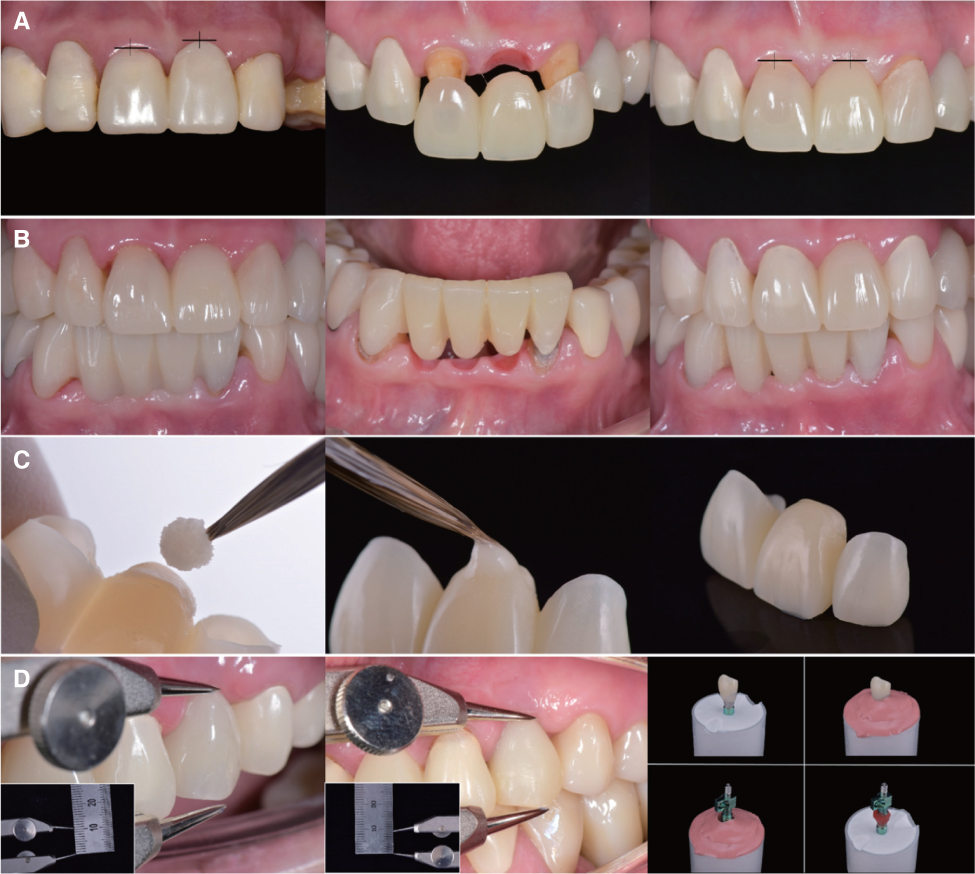
Fig. 7 Adjustment process of anterior provisional restoration. (A) The process of fitting the cervical line of the central anterior teeth using ovate pontic (#21), (B) The pink esthetic of the mandibular incisor obtained by adjusting the countour of cervical embrassure and using ovate pontic, (C) ovate pontic adjustment process at the chair side, (D) The process of fabricating customized pick-up copings after gingival molding to adjust the cervical line of the canine (#23) where implant installed.

Fig. 8 Definitive prosthesis design procedure. (A, D) Master cast and its scanned data, (B, E) Model of provisional restoration and its scanned data, (C, F) Definitive prosthesis fabricated by double scan technique.

Fig. 9 Postoperative intraoral photographs, radiographic evaluation and night guard. (A) Post-operative panoramic radiograph, (B) Maxillary occlusal view, (C) 3D printed night guard, (D) Right lateral view, (E) Frontal view, (F) Left lateral view, (G) Canine guidance at right side, (H) Mandibular occlusal view, (I) Group function at left side.

Fig. 10 Distance between zenith of canine is increased 2.5 mm and It was maintained without change during treatment procedure. (A) Distance between zenith of canine is 17.5 mm before rehabilitation of vertical dimension, (B) Distance between zenith of canine is 20.0 mm at newly determined centric relation, (C) Distance between zenith of canine is 20.0 mm at provisional restoration, (D) Distance between zenith of canine is 20.0 mm at final restoration.
Reference
-
1. Pinho T, Neves M, Alves C. Multidisciplinary management including periodontics, orthodontics, implants, and prosthetics for an adult. Am J Orthod Dentofacial Orthop. 2012; 142:235–245.
Article2. Padwa BL, Kaiser MO, Kaban LB. Occlusal cant in the frontal plane as a reflection of facial asymmetry. J Oral Maxillofac Surg. 1997; 55:811–816.
Article3. Ramfjord SP, Blankenship JR. Increased occlusal vertical dimension in adult monkeys. J Prosthet Dent. 1981; 45:74–83.
Article4. Dawson PE. Functional occlusion: From TMJ to smile design. St. Louis: Mosby Elsevier;2007.5. Hemmings KW, Darbar UR, Vaughan S. Tooth wear treated with direct composite restorations at an increased vertical dimension: results at 30 months. J Prosthet Dent. 2000; 83:287–293.
Article6. Sato S, Hotta TH, Pedrazzi V. Removable occlusal overlay splint in the management of tooth wear: a clinical report. J Prosthet Dent. 2000; 83:392–395.
Article7. Smith BG, Knight JK. A comparison of patterns of tooth wear with aetiological factors. Br Dent J. 1984; 157:16–19.
Article8. Davies SJ, Gray RJ, Qualtrough AJ. Management of tooth surface loss. Br Dent J. 2002; 192:11–16.
Article9. Mohindra NK, Bulman JS. The effect of increasing vertical dimension of occlusion on facial aesthetics. Br Dent J. 2002; 192:164–168.
Article10. Muts EJ, van Pelt H, Edelhoff D, Krejci I, Cune M. Tooth wear: a systematic review of treatment options. J Prosthet Dent. 2014; 112:752–759.11. Lee RL, Gregory GG. Gaining vertical dimension for the deep bite restorative patient. Dent Clin North Am. 1971; 15:743–763.12. Misch CE. Contemporary implant dentistry. 3rd ed. St. Louis: Mosby;2008.13. Widmann G, Bale RJ. Accuracy in computer-aided implant surgery-a review. Int J Oral Maxillofac Implants. 2006; 21:305–313.14. Abboud M, Wahl G, Calvo-Guirado JL, Orentlicher G. Application and success of two stereolithographic surgical guide systems for implant placement with immediate loading. Int J Oral Maxillofac Implants. 2012; 27:634–643.15. Abduo J, Lyons K. Clinical considerations for increasing occlusal vertical dimension: a review. Aust Dent J. 2012; 57:2–10.
Article16. Azari A, Nikzad S. Computer-assisted implantology: historical background and potential outcomes-a review. Int J Med Robot. 2008; 4:95–104.
Article17. Raico Gallardo YN, da Silva-Olivio IRT, Mukai E, Morimoto S, Sesma N, Cordaro L. Accuracy comparison of guided surgery for dental implants according to the tissue of support: a systematic review and meta-analysis. Clin Oral Implants Res. 2017; 28:602–612.
Article18. Schneider D, Schober F, Grohmann P, Hammerle CH, Jung RE. In-vitro evaluation of the tolerance of surgical instruments in templates for computer-assisted guided implantology produced by 3-D printing. Clin Oral Implants Res. 2015; 26:320–325.
Article19. Karl M, Graef F, Wichmann M, Krafft T. Passivity of fit of CAD/CAM and copy-milled frameworks, veneered frameworks, and anatomically contoured, zirconia ceramic, implantsupported fixed prostheses. J Prosthet Dent. 2012; 107:232–238.
Article20. Kim Y, Oh TJ, Misch CE, Wang HL. Occlusal considerations in implant therapy: clinical guidelines with biomechanical rationale. Clin Oral Implants Res. 2005; 16:26–35.
Article
- Full Text Links
-
- Actions
-
Cited
- CITED
-
- Close
- Share
-
- Similar articles
-
- Full-mouth rehabilitation with increasing minimum vertical dimension in the patient with severely worn dentition and deep bite
- Full mouth rehabilitation with vertical dimension increase in patient with loss of anterior guidance due to maxillary anterior teeth wear: A case report
- Full mouth rehabilitation of a patient with worn dentition and loss of posterior support by vertical dimension reestablishment: a clinical report
- Full-mouth rehabilitation without changing the vertical dimension in patient with worn dentition
- Full mouth rehabilitation with extremely worn dentition
