Full mouth rehabilitation using zirconia crown in severe worn dentition: a case report
- Affiliations
-
- 1Department of Prosthodontics, School of Dentistry, Kyungpook National University, Daegu, Republic of Korea. sacho@knu.ac.kr
- KMID: 2357848
- DOI: http://doi.org/10.14368/jdras.2016.32.3.202
Abstract
- The progressive attrition of teeth is a normal process by aging. However, excessive tooth wear with decreased vertical dimension of occlusion and collapse of occlusal plane may cause pathologic pulpal condition, occlusal disharmony and functional disorders. In this case, a patient with severely worn dentition was treated. Diagnostic wax-up was performed at the increased vertical dimension. After evaluation of provisional restorations for 12 weeks, final restorations were fabricated with zirconia crown and routine clinical assessments were made. Esthetically and functionally satisfactory results were obtained.
MeSH Terms
Figure
-
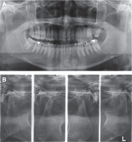
Fig. 1 (A) Panoramic view, (B) TMJ series before treatment. No evidence of pathologic change was seen. Centric occlusion and maximum opening.

Fig. 2 Initial intraoral view. (A) Maxillary occlusal view, (B) Right lateral view, (C) Frontal view, (D) Left lateral view, (E) Mandibular occlusal view.
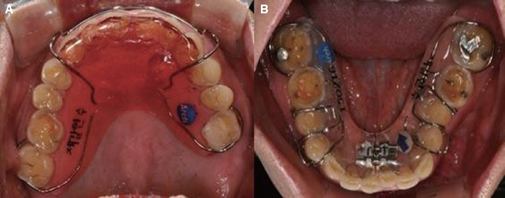
Fig. 3 Orthodontic treatment. (A) Bite plate on maxilla, (B) Schwarts on mandible.
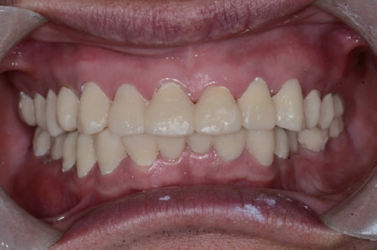
Fig. 4 Temporary prosthesis.
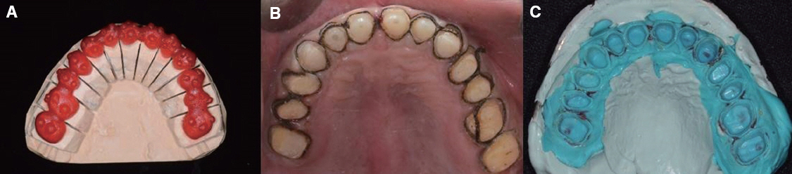
Fig. 5 Maxilla. (A) Making tooth tray, (B) Teeth preparation and code packing, (C) Final impression taking.
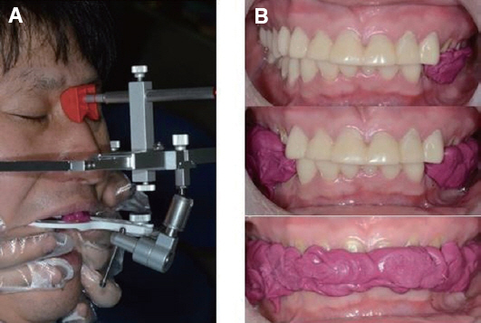
Fig. 6 (A) Facebow transfer, (B) Bite registration.

Fig. 7 Maser cast. (A) Maxillary occlusal view, (B) Right lateral view, (C) Frontal view, (D) Left lateral view, (E) Mandibular occlusal view.
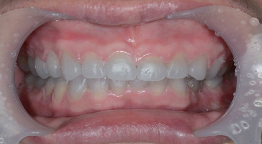
Fig. 8 Milling wax try-in.

Fig. 9 Definitive prosthesis. (A) Maxillary occlusal view, (B) Right lateral view, (C) Frontal view, (D) Left lateral view, (E) Right working movement, (F) Mandibular occlusal view, (G) Left working movement.
Reference
-
References
1. Verrett RG. Analyzing the etiology of an extremely worn dentition. J Prosthodont. 2001; 10:224–33. DOI: 10.1053/jpro.2001.28264. PMID: 11781971.2. Dawson PE. Functional occlusion - from TMJ to smile design. 1st ed. St. Louis: Mosby;2006. p. 430–3.3. Turner KA, Missirlian DM. Restoration of the extremely worn dentition. J Prosthet Dent. 1984; 52:467–74. DOI: 10.1016/0022-3913(84)90326-3.4. Ramfjord SP. Bruxism, a clinical and electromyographic study. J Am Dent Assoc. 1961; 62:21–44. DOI: 10.14219/jada.archive.1961.0002. PMID: 13739329.5. Carlsson GE, Ingervall B, Kocak G. Effect of increasing vertical dimension on the masticatory system in subjects with natural teeth. J Prosthet Dent. 1979; 41:284–9. DOI: 10.1016/0022-3913(79)90008-8.6. Albashaireh ZS, Ghazal M, Kern M. Two-body wear of different ceramic materials opposed to zirconia ceramic. J Prosthet Dent. 2010; 104:105–13. DOI: 10.1016/S0022-3913(10)60102-3.
- Full Text Links
-
- Actions
-
Cited
- CITED
-
- Close
- Share
-
- Similar articles
-
- Full mouth rehabilitation with vertical dimension increase in patient with severly worn dentition
- Full mouth rehabilitation of a patient with worn dentition and loss of posterior support by vertical dimension reestablishment: a clinical report
- Full-mouth rehabilitation without changing the vertical dimension in patient with worn dentition
- Oral rehabilitation of a patient with severely worn dentition using monolithic zirconia
- Full mouth rehabilitation of the patient with worn dentition using full-contour monolithic zirconia prostheses at an increased vertical dimension of occlusion: a case report
