Osteochondroma at the Cervicothoracic Junction
- Affiliations
-
- 1Department of Orthopedic Surgery, National Health Insurance Service Ilsan Hospital, Goyang, Korea. hjwspine@gmail.com
- 2Department of Pathology, National Health Insurance Service Ilsan Hospital, Goyang, Korea.
- KMID: 2469888
- DOI: http://doi.org/10.4055/jkoa.2019.54.6.562
Abstract
- Osteochondroma is a common benign bone tumor, but relatively rare in the spine. Spinal osteochondroma originates mainly from the posterior column and sometimes invades the spinal canal causing cord compression and myelopathy. A 36-year-old man was admitted to the authors' institution with a two-year history of neck pain. A radiology examination revealed osteochondroma, arising from the left superior articular process of the T1 vertebra with significant central canal narrowing. Most cases of the spinal osteochondroma originate in the cervical vertebra, but there are few reports of an origin of the cervicothoracic junction. This paper reports a case of osteochondroma at the cervicothoracic junction with a review of the relevant literature.
Keyword
Figure
-
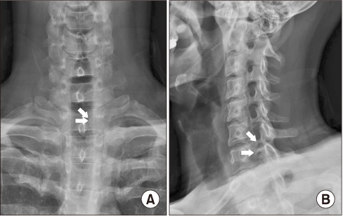
Figure 1 (A) Simple anteroposterior radiograph showing a bulging contour with left face joint hypertrophy on the C-T junction (arrows). (B) Left oblique radiograph showing C7/T1 foramen stenosis by facet joint hypertrophy (arrows).
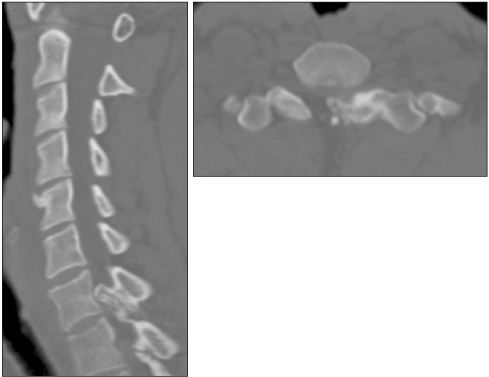
Figure 2 Computed tomography showing a 1.3×0.9×1.0 cm size bony mass arising from the left superior articular process of the T1 vertebra with significant central canal narrowing.
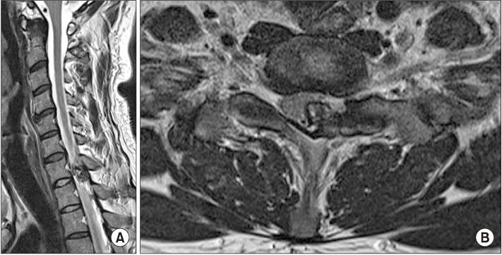
Figure 3 Magnetic resonance imaging showing osteochondroma arising from the T1 left superior articular process causing cord compression (A) and equivocal cord signal changes (B).
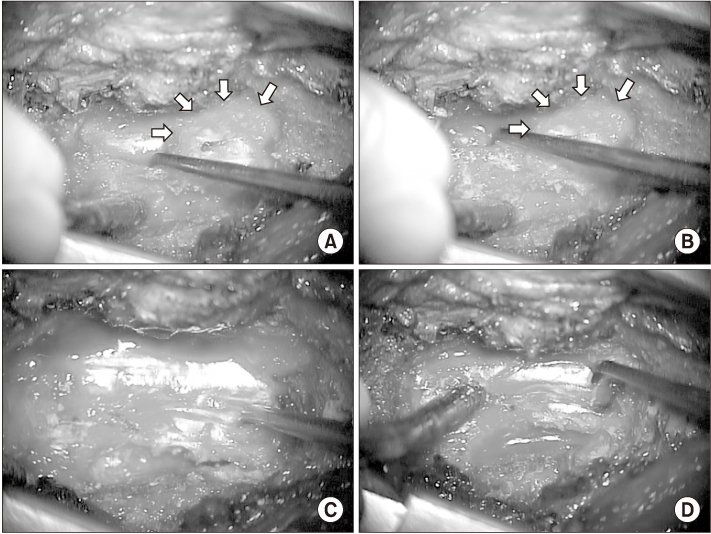
Figure 4 Partial laminectomy and microscopic en-bloc mass excision was performed on the patient. (A, B) Protruded mass (arrows). (C, D) After excision.
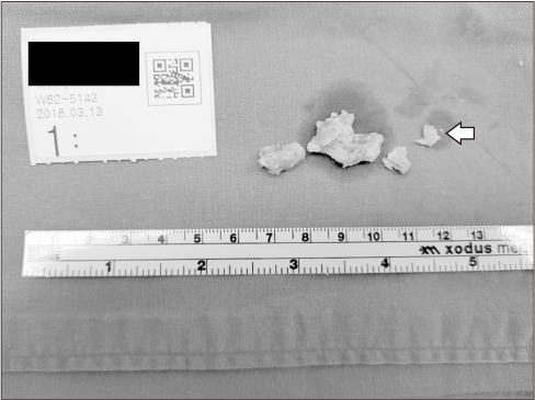
Figure 5 Gross photograph of the removed mass (arrow) and laminae.
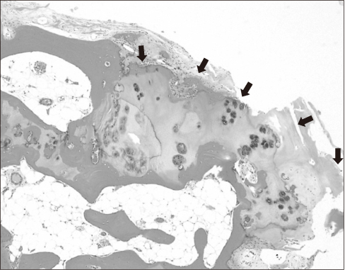
Figure 6 Histologically, cap (arrows) composed of mature hyaline cartilage with overlying fibrous perichondrium is observed over the linearly arranged cancellous bone and fatty marrow (H&E, ×100). Scattered enchondral ossification is noted.
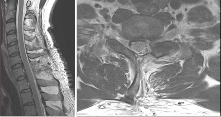
Figure 7 On postoperative magnetic resonance imaging, the osteochondroma was removed and the spinal cord was decompressed.
Reference
-
1. Sugiyama H, Omonishi K, Yonehara S, et al. Characteristics of benign and malignant bone tumors registered in the hiroshima tumor tissue registry, 1973-2012. JB JS Open Access. 2018; 3:e0064.
Article2. Yakkanti R, Onyekwelu I, Carreon LY, Dimar JR 2nd. Solitary osteochondroma of the spine-a case series: review of solitary osteochondroma with myelopathic symptoms. Global Spine J. 2018; 8:323–339.
Article3. O'Brien MF, Bridwell KH, Lenke LG, Schoenecker PL. Intracanalicular osteochondroma producing spinal cord compression in hereditary multiple exostoses. J Spinal Disord. 1994; 7:236–241.4. Sinelnikov A, Kale H. Osteochondromas of the spine. Clin Radiol. 2014; 69:e584–e590.
Article5. Chatzidakis E, Lypiridis S, Kazdaglis G, Chatzikonstadinou K, Papatheodorou G. A rare case of solitary osteochondroma of the dens of the C2 vertebra. Acta Neurochir (Wien). 2007; 149:637–638.
Article6. Ratliff J, Voorhies R. Osteochondroma of the C5 lamina with cord compression: case report and review of the literature. Spine (Phila Pa 1976). 2000; 25:1293–1295.7. Jang JH, Cho ST, Son JM, Ha NK, Song ES, Yang YJ. Osteochondroma of the 5th and 6th cervical vertebral body: one case report. J Korean Soc Spine Surg. 2005; 12:238–244.8. Arasil E, Erdem A, Yüceer N. Osteochondroma of the upper cervical spine. A case report. Spine (Phila Pa 1976). 1996; 21:516–518.
- Full Text Links
-
- Actions
-
Cited
- CITED
-
- Close
- Share
-
- Similar articles
-
- Fractures and Dislocations of the Cervicothoracic Junction
- Cervicothoracic Junction Approach using Modified Anterior Approach: J-type Manubriotomy and Low Cervical Incision
- Osteochondroma of the Talus: A Report of Two Cases
- Migrated Disc at Cervicothoracic Junction Presenting as Acute Paraplegia
- A Case of Osteochondroma which Arised form Right Side lamina of 5th Lumbar Vertebra
