Ann Dermatol.
2020 Feb;32(1):14-20. 10.5021/ad.2020.32.1.14.
Mycosis Fungoides and Variants of Mycosis Fungoides: A Retrospective Study of 93 Patients in a Chinese Population at a Single Center
- Affiliations
-
- 1Department of Dermatology, Peking Union Medical College Hospital, Chinese Academy of Medical Sciences and Peking Union Medical College, Beijing, China. liujie04672@pumch.cn, yuehualiu@263.net
- 2Department of Hematology, Peking Union Medical College Hospital, Chinese Academy of Medical Sciences and Peking Union Medical College, Beijing, China.
- KMID: 2467196
- DOI: http://doi.org/10.5021/ad.2020.32.1.14
Abstract
- BACKGROUND
Mycosis fungoides (MF) is the most common types of cutaneous T cell lymphoma. It typically presents with erythematous patches and plaques in the early stage and tumors and extracutaneous involvement with possibly fatal outcomes in the late stage. To facilitate early and accurate diagnosis of MF, it is essential to be knowledgeable of classic and variants of this disease. However, there is limited published data in the Chinese population.
OBJECTIVE
To characterize our patient group and to provide additional insight into these malignancies.
METHODS
Patients diagnosed with mycosis fungoides and its variants from October 2012 to January 2018 were retrospectively analyzed. Disease-specific survival (DSS) rate and curve according to early and advanced stages were also calculated.
RESULTS
The mean age at diagnosis of ninety-three MF patients was 38.9±1.73 years (range: 6~77). Forty-five males (48.4%) and 48 females (51.6 %) were included in this study. The DSS rate of early-stage MF was 98.6%, while that of advanced stage MF was 88.9%. There was a significant difference in DSS rate between early stage and advanced stage MF (p=00.042, log-rank test). The median age of 10 patients with hypopigmented MF (hMF) was 10.5 years (range: 6~28). The age of hMF was younger than that of classical MF patients (p < 0.05).
CONCLUSION
Early-stage MF has a better prognosis than advanced stage and hMF affects younger people than classic MF among Chinese. This study provides an insight into mycosis fungoides and its variants in a Chinese population.
Keyword
MeSH Terms
Figure
-
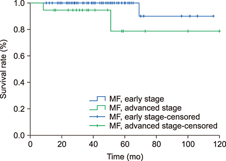
Fig. 1 Disease-specific survival curve according to early-stage mycosis fungoides (MF) and advanced-stage MF.
Reference
-
1. Willemze R, Jaffe ES, Burg G, Cerroni L, Berti E, Swerdlow SH, et al. WHO-EORTC classification for cutaneous lymphomas. Blood. 2005; 105:3768–3785.
Article2. Souissi A, Ben Lagha I, Jendoubi F, Drissi H, Chelly I, Mokni M. Spiky follicular mycosis fungoides: a trichoscopic feature. J Eur Acad Dermatol Venereol. 2019; 33:e252–e253.
Article3. Rodney IJ, Kindred C, Angra K, Qutub ON, Villanueva AR, Halder RM. Hypopigmented mycosis fungoides: a retrospective clinicohistopathologic study. J Eur Acad Dermatol Venereol. 2017; 31:808–814.
Article4. Pankratov O, Gradova S, Tarasevich S, Pankratov V. Poikilodermatous mycosis fungoides: clinical and histopathological analysis of a case and literature review. Acta Dermatovenerol Alp Pannonica Adriat. 2015; 24:37–41.
Article5. Willemze R, Cerroni L, Kempf W, Berti E, Facchetti F, Swerdlow SH, et al. The 2018 update of the WHO-EORTC classification for primary cutaneous lymphomas. Blood. 2019; 133:1703–1714.
Article6. Ku LS, Lo KK. Mycosis fungoides--a retrospective study of 40 cases in Hong Kong. Int J Dermatol. 2005; 44:215–220.7. Ishiji T, Takagi Y, Niimura M. Cutaneous lymphomas in Tokyo: analysis of 62 cases in a dermatology clinic. Int J Dermatol. 2001; 40:37–40.
Article8. Liu J, Yu X, Liu Y, Jin H, Ma D, Qu T, et al. Relative frequency and survival of primary cutaneous lymphomas: a retrospective analysis of 98 patients. Chin Med J (Engl). 2014; 127:645–650.9. Tan EST, Tang MBY, Tan SH. Retrospective 5XMLLink_XYZyear review of 131 patients with mycosis fungoides and Sézary syndrome seen at the National Skin Centre, Singapore. Australas J Dermatol. 2006; 47:248–252.
Article10. Su C, Nguyen KA, Bai HX, Cao Y, Tao Y, Xiao R, et al. Racial disparity in mycosis fungoides: an analysis of 4495 cases from the US National Cancer Database. J Am Acad Dermatol. 2017; 77:497–502.e2.
Article11. Eder J, Kern A, Moser J, Kitzwögerer M, Sedivy R, Trautinger F. Frequency of primary cutaneous lymphoma variants in Austria: retrospective data from a dermatology referral centre between 2006 and 2013. J Eur Acad Dermatol Venereol. 2015; 29:1517–1523.
Article12. Bradford PT, Devesa SS, Anderson WF, Toro JR. Cutaneous lymphoma incidence patterns in the United States: a population-based study of 3884 cases. Blood. 2009; 113:5064–5073.
Article13. Agar NS, Wedgeworth E, Crichton S, Mitchell TJ, Cox M, Ferreira S, et al. Survival outcomes and prognostic factors in mycosis fungoides/Sézary syndrome: validation of the revised International Society for Cutaneous Lymphomas/European Organisation for Research and Treatment of Cancer staging proposal. J Clin Oncol. 2010; 28:4730–4739.
Article14. Lebowitz E, Geller S, Flores E, Pulitzer M, Horwitz S, Moskowitz A, et al. Survival, disease progression and prognostic factors in elderly patients with mycosis fungoides and Sézary syndrome: a retrospective analysis of 174 patients. J Eur Acad Dermatol Venereol. 2019; 33:108–114.
Article15. Olsen EA, Hodak E, Anderson T, Carter JB, Henderson M, Cooper K, et al. Guidelines for phototherapy of mycosis fungoides and Sézary syndrome: a consensus statement of the United States Cutaneous Lymphoma Consortium. J Am Acad Dermatol. 2016; 74:27–58.
Article16. Stadler R, Otto HG, Luger T, Henz BM, Kuhl P, Zwingers T, et al. Prosepctive randomized multicenter clinical traial on the use of interferon-2α plus acitretin Photo(chemo)therapie April 2007 24 versus interferon - 2α plus PUVA in patients with cutaneous T-cell lymphoma stages I and II. Blood. 1998; 92:3578–3581.17. Olisova OY, Megna M, Grekova EV, Zaslavsky DV, Gorenkova LG, Sidikov AA, et al. PUVA and interferon α2b combined therapy for patients with mycosis fungoides at different stages of the disease: a seven-year retrospective study in Russia. J Eur Acad Dermatol Venereol. 2019; 33:e72–e74.
Article18. van Doorn R, Van Haselen CW, van Voorst Vader PC, Geerts ML, Heule F, de Rie M, et al. Mycosis fungoides: disease evolution and prognosis of 309 Dutch patients. Arch Dermatol. 2000; 136:504–510.19. Lee HS, Suh KS, Lee DY, Cho KH, Oh SH, Kim SC, et al. Cutaneous lymphoma in Korea: a nationwide retrospective study. Acta Derm Venereol. 2016; 96:535–539.
Article20. Park JH, Shin HT, Lee DY, Lee JH, Yang JM, Jang KT, et al. World Health Organization-European Organization for Research and Treatment of Cancer classification of cutaneous lymphoma in Korea: a retrospective study at a single tertiary institution. J Am Acad Dermatol. 2012; 67:1200–1209.
Article21. Abeldaño A, Enz P, Maskin M, Cervini AB, Torres N, Acosta AC, et al. Primary cutaneous lymphoma in Argentina: a report of a nationwide study of 416 patients. Int J Dermatol. 2019; 58:449–455.
Article22. van Santen S, van Doorn R, Neelis KJ, Daniëls LA, Horváth B, Bruijn MS, et al. Recommendations for treatment in folliculotropic mycosis fungoides: report of the Dutch Cutaneous Lymphoma Group. Br J Dermatol. 2017; 177:223–228.
Article23. Kempf W, Ostheeren-Michaelis S, Paulli M, Lucioni M, Wechsler J, Audring H, et al. Cutaneous Lymphoma Histopathology Task Force Group of the European Organization for Research and Treatment of Cancer. Granulomatous mycosis fungoides and granulomatous slack skin: a multicenter study of the Cutaneous Lymphoma Histopathology Task Force Group of the European Organization For Research and Treatment of Cancer (EORTC). Arch Dermatol. 2008; 144:1609–1617.
Article24. Amorim GM, Niemeyer-Corbellini JP, Quintella DC, Cuzzi T, Ramos-E-Silva M. Hypopigmented mycosis fungoides: a 20-case retrospective series. Int J Dermatol. 2018; 57:306–312.
Article25. Ardigó M, Borroni G, Muscardin L, Kerl H, Cerroni L. Hypopigmented mycosis fungoides in Caucasian patients: a clinicopathologic study of 7 cases. J Am Acad Dermatol. 2003; 49:264–270.
Article26. Boulos S, Vaid R, Aladily TN, Ivan DS, Talpur R, Duvic M. Clinical presentation, immunopathology, and treatment of juvenile-onset mycosis fungoides: a case series of 34 patients. J Am Acad Dermatol. 2014; 71:1117–1126.
Article
