Clin Endosc.
2019 Nov;52(6):624-625. 10.5946/ce.2019.083.
Delayed Duodenal Perforation of an Endoscopic Mucosal Resection-Induced Ulcer due to a Foreign Body
- Affiliations
-
- 1Division of Gastroenterology, Department of Internal Medicine, College of Medicine, Incheon St. Mary’s Hospital, The Catholic University of Korea, Seoul, Korea. huhcw@catholic.ac.kr
- KMID: 2465808
- DOI: http://doi.org/10.5946/ce.2019.083
Abstract
- No abstract available.
MeSH Terms
Figure
-
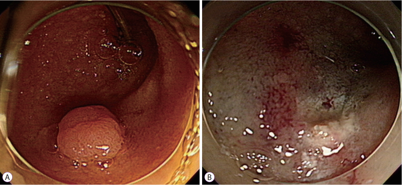
Fig. 1. (A) An esophagogastroduodenoscopy image of a subtly yellowish, elevated lesion 10 mm in diameter in the afferent loop of a Billroth II anastomosis. (B) Clear resection site after endoscopic mucosal resection.

Fig. 2. Images after 2 days. (A) A computed tomography scan showing fluid collection and free air around the afferent loop and right colon (yellow arrow). (B, C) An esophagogastroduodenoscopy image showing a sharp foreign body penetrating the endoscopic mucosal resection site. (D) The foreign body is a suture material used in the previous gastric surgery.
Reference
-
1. Yahagi N, Kato M, Ochiai Y, et al. Outcomes of endoscopic resection for superficial duodenal epithelial neoplasia. Gastrointest Endosc. 2018; 88:676–682.
Article2. Tomizawa Y, Ginsberg GG. Clinical outcome of EMR of sporadic, nonampullary, duodenal adenomas: a 10-year retrospective. Gastrointest Endosc. 2018; 87:1270–1278.
Article3. Ochiai Y, Kato M, Kiguchi Y, et al. Current status and challenges of endoscopic treatments for duodenal tumors. Digestion. 2019; 99:21–26.
Article4. Kato M, Ochiai Y, Fukuhara S, et al. Clinical impact of closure of the mucosal defect after duodenal endoscopic submucosal dissection. Gastrointest Endosc. 2019; 89:87–93.
Article
- Full Text Links
-
- Actions
-
Cited
- CITED
-
- Close
- Share
-
- Similar articles
-
- Endoscopic Suturing for the Prevention and Treatment of Complications Associated with Endoscopic Mucosal Resection of Large Duodenal Adenomas
- Liver Abscess Secondary to Perforation after Duodenal Endoscopic Resection
- Two Cases of Successful Clipping Closure of Iatrogenic Duodenal Perforation Occurred during Endoscopic Procedure
- Endoscopic Clip Ligation on Mucosal Defect after Endoscopic Mucosal Resection
- Endoscopic Removal of Esophageal Foreign Body Complicated with Esophageal Ulcer: Case report
