J Korean Neurosurg Soc.
2019 Nov;62(6):723-726. 10.3340/jkns.2018.0225.
Spontaneously Regressed Rathke's Cleft Cyst
- Affiliations
-
- 1Department of Neurosurgery, School of Medicine, Kyungpook National University, Daegu, Korea. nsdoctor@naver.com
- 2Department of Neurosurgery, Kyungpook National University Hospital, Daegu, Korea.
- KMID: 2463664
- DOI: http://doi.org/10.3340/jkns.2018.0225
Abstract
- We report two rare cases of spontaneously regressed Rathke's cleft cyst (RCC). A 52-year-old woman presented with headache. A pituitary hormone study was normal. Brain magnetic resonance imaging (MRI) showed a 0.45-cm³ cystic sellar lesion. The cyst was hyperintense on T1-weighed imaging and hypointense on T2-weighted imaging without rim enhancement, comparable to a RCC. Six months later, brain MRI showed no change in the cyst size. Without any medical treatments, brain MRI 1 year later revealed a spontaneous decrease in cyst volume to 0.05 cm³. A 34-year-old woman presented with headache and galactorrhea lasting 1 week. At the time of the visit, the patient's headache had disappeared. Her initial serum prolactin level was 81.1 ng/mL, and after 1 week without the cold medicine, the serum prolactin level normalized to 11.28 ng/mL. Brain MRI showed a RCC measuring 0.71 cm³. Without further treatments, brain computed tomography 6 months later showed a spontaneous decrease in cyst volume to 0.07 cm³. Another 6 months later, brain MRI revealed that the cyst had remained the same size. Neither patient experienced neurological symptoms, such as headache or visual disturbance, during the period of cyst reduction. The RCCs in both patients underwent spontaneous regression without any medical treatment during a period of 6 months to 1 year. Although spontaneous regression of a RCC is rare, it is still possible and a sufficient follow-up period should be considered.
MeSH Terms
Figure
-
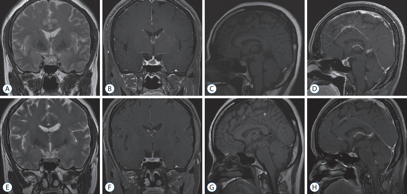
Fig. 1. Brain magnetic resonance imaging (MRI) revealing a Rathke’s cleft cyst measuing 0.45 cm3 (A-D). Subsequent brain MRI revealing a spontaneous decrease in volume to 0.05 cm3, 1 year later (E-H).
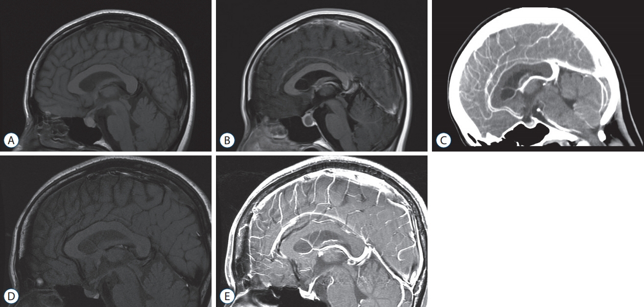
Fig. 2. Brain magnetic resonance imaging (MRI) revealing a Rathke’s cleft cyst measuring 0.71 cm3 (A and B). Six months later, brain computed tomography showing the spontaneous decrease in volume to 0.07 cm3 (C). Another six months later, brain MRI showing no change in size (D and E).
Reference
-
References
1. Aho CJ, Liu C, Zelman V, Couldwell WT, Weiss MH. Surgical outcomes in 118 patients with Rathke cleft cysts. J Neursurg. 102:189–193. 2005.
Article2. Al Safatli D, Kalff R, Waschke A. Spontaneous involution of a presumably Rathke’s cleft cyst in a patient with slight subclinical hypopituitarism: a case report and review of the literature. Case Rep Surg. 2015:971364–971365. 2015.
Article3. Amhaz HH, Chamoun RB, Waguespack SG, Shah K, McCutcheon IE. Spontaneous involution of Rathke cleft cysts: is it rare or just underreported? J Neurosurg. 112:1327–1332. 2010.
Article4. Cheng L, Guo P, Jin P, Li H, Fan M, Cai E. Spontaneous involution of a Rathke cleft cyst. J Craniofac Surg. 27:e791–e793. 2016.
Article5. Hatayama K, Abe T, Endo T, Kobayashi N, Shimazu M, Sasaki K, et al. Pituitary cystic mass with spontaneous disappearance: three cases of equivocal Rathke’s cleft cyst. No To Shinkei. 52:929–933. 2000.6. Igarashi T, Saeki N, Yamaura A. Long-term magnetic resonance imaging follow-up of asymptomatic sellar tumors. Their natural history and surgical indications. Neurol Med Chir (Tokyo). 39:592–598. 1999.
Article7. Kim CW, Hwang K, Joo JD, Kim YH, Han JH, Kim CY. Spontaneous involution of Rathke’s cleft cysts without visual symptoms. Brain Tumor Res Treat. 4:58–62. 2016.
Article8. Maruyama H, Iwasaki Y, Tsugita M, Ogami N, Asaba K, Takao T, et al. Rathke’s cleft cyst with short-term size changes in response to glucocorticoid replacement. Endocr J. 55:425–428. 2008.
Article9. Munich SA, Leonardo J. Spontaneous involution of a Rathke’s cleft cyst in a patient with normal cortisol secretion. Surg Neurol Int. 3:42. 2012.
Article10. Nishio S, Morioka T, Suzuki S, Fukui M. Spontaneous regression of a pituitary cyst: report of two cases. Clin Imaging. 25:15–17. 2001.
Article11. Rasmussen Z, Abode-Iyamah KO, Kirby P, Greenlee JD. Rathke’s cleft cyst: a case report of recurrence and spontaneous involution. J Clin Neurosci. 32:122–125. 2016.
Article12. Saeki N, Sunami K, Sugaya Y, Yamaura A. MRI findings and clinical manifestations in Rathke’s cleft cyst. Acta Neurochir (Wien). 141:1055–1061. 1999.
Article13. Simmons JD, Simmons LA. Spontaneous regression of a pituitary cyst. Neuroradiology. 41:27–29. 1999.
Article
- Full Text Links
-
- Actions
-
Cited
- CITED
-
- Close
- Share
-
- Similar articles
-
- Symptomatic Rathke's Cleft Cyst in the Interpeduncular Cistern: Case Report
- Suprasellar Rathke Cleft Cyst: A case report
- Rathke's Cleft Cyst: Case Report
- Large Ossified Rathke's Cleft Cyst: A Case Report and Review of the Literature
- Pituitary Tumors Composed of Adenohypophysial Adenoma and Rathke's Cleft Cyst Elements
