Malignant Hepatic Solitary Fibrous Tumor
- Affiliations
-
- 1Department of Plastic and Reconstructive Surgery, Kosin University College of Medicine, Busan, Korea. hose3290@naver.com
- 2Department of Internal Medicine, Gupo Sungshim Hospital, Busan, Korea.
- KMID: 2463615
- DOI: http://doi.org/10.17998/jlc.19.2.143
Abstract
- Hepatic solitary fibrous tumors (SFTs) are mostly benign and rare because of information regarding the clinical symptoms, treatment, and prognosis of their malignant forms is currently lacking. A literature review concerning malignant SFTs revealed that there were a few cases where patients experienced abdominal right upper quadrant (RUQ) pain as their first clinical symptom, and metastases were found after being diagnosed with hepatic SFT. Here, we report a patient who was previously healthy without any clinical symptoms such as RUQ pain or weight loss, but had the appearance of a metastatic mass as the first clinical presentation before a primary hepatic SFT was detected.
Keyword
MeSH Terms
Figure
-
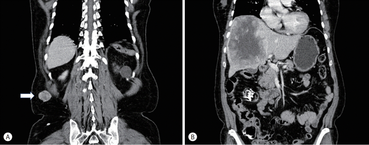
Figure 1. Abdominal computed tomography coronal view. (A) Heterogeneous intermediate density enhanced mass was observed in the right flank region (arrow). (B) An approximately 14×11×10 cm sized mass was accidentally detected in the liver.
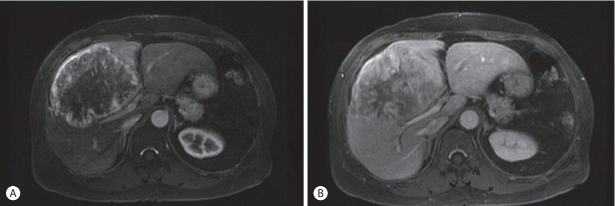
Figure 2. Liver third phase magnetic resonance imaging axial view. Approximately 14×11×10 cm sized large intermediate signal intensity mass, peripheral enhanced rim with internal necrotic portion, peritumor bile duct dilatations from segment IV of the liver to the right hepatic lobe. (A) Arterial phase: early arterial enhancement. (B) Portal phase: delayed progress enhancement findings.
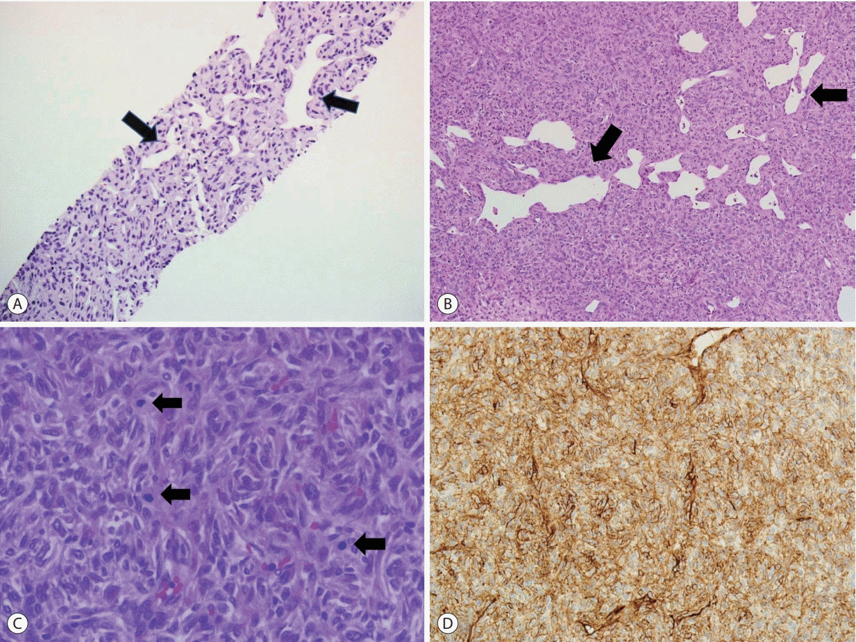
Figure 3. Histological findings. (A) Histology of the liver shows similar morphology to spindled tumor cells with a typical “staghorn” vascular pattern (arrows) (hematoxylin and eosin staining [H&E], ×100). (B) Histology of the right flank mass. The tumor cells are arranged in a “patternless” disposition with typical ectatic and irregularly shaped vessels (arrows) (H&E, ×100). (C) Histology of the right flank mass. At high power, tumors are ovoid to spindle shaped with relatively uniform and bland cytology. Increased mitotic activity (arrows) is identified (H&E, ×400). (D) CD34 immunohistochemical expression is strong and diffuse (×200).
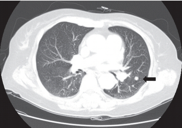
Figure 4. Chest computed tomography findings with a small nodule in the left lower lobe of the lung, which was suspected to be metastasis (arrow).
Reference
-
1. Briselli M, Mark EJ, Dickersin GR. Solitary fibrous tumors of the pleura: eight new cases and review of 360 cases in the literature. Cancer. 1981; 47:2678–2689.2. Hasegawa T, Matsuno Y, Shimoda T, Hasegawa F, Sano T, Hirohashi S. Extrathoracic solitary fibrous tumors: their histological variability and potentially aggressive behavior. Hum Pathol. 1999; 30:1464–1473.3. Perini MV, Herman P, D’Albuquerque LA, Saad WA. Solitary fibrous tumor of the liver: report of a rare case and review of the literature. Int J Surg. 2008; 6:396–399.4. Yilmaz S, Kirimlioglu V, Ertas E, Hilmioglu F, Yildirim B, Katz D, et al. Giant solitary fibrous tumor of the liver with metastasis to the skeletal system successfully treated with trisegmentectomy. Dig Dis Sci. 2000; 45:168–174.5. Jakob M, Schneider M, Hoeller I, Laffer U, Kaderli R. Malignant solitary fibrous tumor involving the liver. World J Gastroenterol. 2013; 19:3354–3357.6. Korkolis DP, Apostolaki K, Aggeli C, Plataniotis G, Gontikakis E, Volanaki D, et al. Solitary fibrous tumor of the liver expressing CD34 and vimentin: a case report. World J Gastroenterol. 2008; 14:6261–6264.7. Archontaki M, Korkolis DP, Arnogiannaki N, Hatzijiannis S, Dendrinos P, Megapanos C, et al. Histologically malignant solitary fibrous tumour of the anterior thoracic wall: a case report and review of the literature. Case Rep Med. 2010; 2010:257167.8. Park MS, Patel SR, Ludwig JA, Trent JC, Conrad CA, Lazar AJ, et al. Activity of temozolomide and bevacizumab in the treatment of locally advanced, recurrent, and metastatic hemangiopericytoma and malignant solitary fibrous tumor. Cancer. 2011; 117:4939–4947.9. Yang X, Zheng J, Ye W, Wang Y, Zhu HG, Wang LZ, et al. Malignant solitary fibrous tumors of the head and neck: a clinicopathological study of nine consecutive patients. Oral Oncol. 2009; 45:678–682.10. Kandpal H, Sharma R, Gupta SD, Kumar A. Solitary fibrous tumour of the liver: a rare imaging diagnosis using MRI and diffusionweighted imaging. Br J Radiol. 2008; 81:e282–e286.11. Fuksbrumer MS, Klimstra D, Panicek DM. Solitary fibrous tumor of the liver: imaging findings. Am J Roentgenol. 2000; 175:1683–1687.12. Bishop JA, Rekhtman N, Chun J, Wakely Jr PE, Ali SZ. Malignant solitary fibrous tumor: cytopathologic findings and differential diagnosis. Cancer Cytopathol. 2010; 118:83–89.
- Full Text Links
-
- Actions
-
Cited
- CITED
-
- Close
- Share
-
- Similar articles
-
- Malignant Solitary Fibrous Tumor of Retroperitoneum Mimicking Gastric Submucosal Tumor
- A Case of Solitary Fibrous Tumor in the Cheek
- Solitary Fibrous Tumor of the Adrenal Gland: A Case Report
- CSF Leakage through a Subarachnoid-pleural Fistula after Resection of a Malignant Solitary Fibrous Tumor
- A Case of Solitary Fibrous Tumor That Developed on the Scalp
