Korean J Gastroenterol.
2011 Jan;57(1):47-50. 10.4166/kjg.2011.57.1.47.
Malignant Solitary Fibrous Tumor of Retroperitoneum Mimicking Gastric Submucosal Tumor
- Affiliations
-
- 1Department of Surgery, Yeungnam University College of Medicine, Daegu, Korea. swkim@med.yu.ac.kr
- KMID: 991469
- DOI: http://doi.org/10.4166/kjg.2011.57.1.47
Abstract
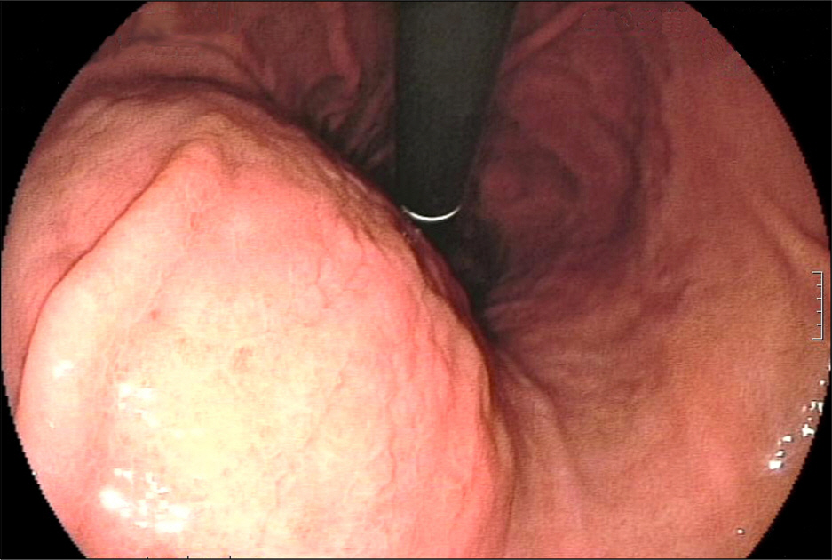
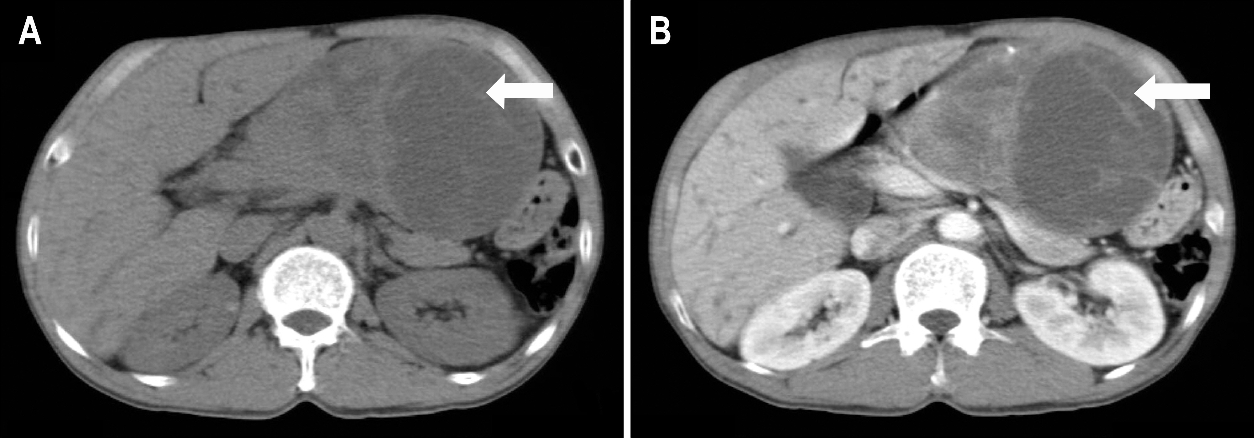
- Solitary fibrous tumors (SFTs) are an uncommon neoplasm characterized by the proliferation of spindle cells. The diagnostic criteria of malignant solitary fibrous tumors (MSFTs) include high cellularity, high mitotic activity (4>10 HPF), pleomorphism, hemorrhage and necrosis. This tumor frequently involves the pleura and MSFTs of retroperitoneum mimicking gastric submucosal tumor are very rare. We report a rare case of MSFT that presented as a gastric submucosal tumor. A gastroscopic examination showed a large bulging mucosa in the gastric body. Abdominal computed tomography revealed a well-defined heterogeneous enhancing mass between the left hepatic lobe and gastric body. Surgical resection was performed and histologic features were consistent with a MSFT.
MeSH Terms
Figure
Reference
-
References
1. England DM, Hochholzer L, McCarthy MJ. Localized benign and malignant fibrous tumors of the pleura. A clinicopathologic review of 223 cases. Am J Surg Pathol. 1989; 13:640–658.2. Young RH, Clement PB, McCaughey WT. Solitary fibrous tumors ('fibrous mesotheliomas') of the peritoneum. A report of three cases and a review of the literature. Arch Pathol Lab Med. 1990; 114:493–495.3. Hasegawa T, Matsuno Y, Shimoda T, Hasegawa F, Sano T, Hirohashi S. Extrathoracic solitary fibrous tumors: their histological variability and potentially aggressive behavior. Hum Pathol. 1999; 30:1464–1473.
Article4. Tanaka M, Sawai H, Okada Y, et al. Malignant solitary fibrous tumor originating from the peritoneum and review of the literature. Med Sci Monit. 2006; 12:CS95–98.5. Cardinale L, Allasia M, Ardissone F, et al. CT features of solitary fibrous tumour of the pleura: experience in 26 patients. Radiol Med. 2006; 111:640–650.
Article6. Hanau CA, Miettinen M. Solitary fibrous tumor: histological and immunohistochemical spectrum of benign and malignant variants presenting at different sites. Hum Pathol. 1995; 26:440–449.
Article7. Chan JK. Solitary fibrous tumour–everywhere, and a diagnosis in vogue. Histopathology. 1997; 31:568–576.8. Kishi K, Homma S, Tanimura S, Matsushita H, Nakata K. Hypoglycemia induced by secretion of high molecular weight insulin-like growth factor-II from a malignant solitary fibrous tumor of the pleura. Intern Med. 2001; 40:341–344.
Article9. Vallat-Decouvelaere AV, Dry SM, Fletcher CD. Atypical and malignant solitary fibrous tumors in extrathoracic locations: evidence of their comparability to intra-thoracic tumors. Am J Surg Pathol. 1998; 22:1501–1511.
- Full Text Links
- Actions
-
Cited
- CITED
-
- Close
- Share
- Similar articles
-
- Myxoid Solitary Fibrous Tumor of the Retroperitoneum: MRI Findings with the Pathologic Correlation
- A Case of the retroperitoneal Malignant Fibrous Histiocytoma
- Pleural Localized Malignant Mesothelioma Mimicking a Benign Solitary Fibrous Tumor of the Pleura on Chest Computed Tomography: A Case Report
- Malignant Solitary Fibrous Tumor in Anterior Mediastinum with Pleural Metastasis Simulating Invasive Thymoma
- Solitary Fibrous Tumor of the Adrenal Gland: A Case Report


