Full mouth rehabilitation with vertical dimension increase in patient with loss of anterior guidance due to maxillary anterior teeth wear: A case report
- Affiliations
-
- 1Department of Prosthodontics, Veteran's Health Medical Service Center, Seoul, Republic of Korea. lysang21@hanmail.net
- KMID: 2377186
- DOI: http://doi.org/10.4047/jkap.2017.55.2.171
Abstract
- Severely worn dentition is frequently multifactorial. It is crucial that the etiology of excessive wear be determined, but accurately diagnosing the factors responsible for tooth wear is often confusing. Before initiating the treatment of these cases, meticulous examination and determining vertical dimension are essential. A 69-year-old male patient had the chief complaint that he has worn dentition and functional and esthetic discomfort. Based on model analysis and diagnostic wax up, new vertical dimension had been determined. Provisional restorations were cemented and after 5 months permanent prostheses were fabricated. This case reports a satisfactory functional and esthetic clinical outcome achieved by restoring the vertical dimension.
MeSH Terms
Figure
-

Fig. 1 Extraoral photograph before treatment. (A) Lateral view, (B) Frontal view.

Fig. 2 Intraoral photograph before treatment. (A) Upper, (B) Right, (C) Frontal, (D) Left, (E) Lower.
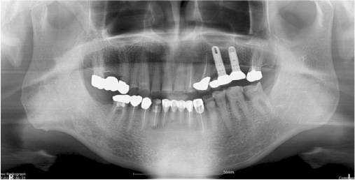
Fig. 3 Panoramic radiograph before treatment.

Fig. 4 Diagnostic wax-up with an increase of 2.0 mm of incisal guide pin.
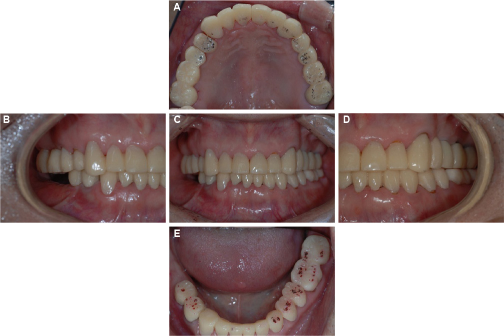
Fig. 5 Provisional prosthesis. (A) Upper, (B) Right, (C) Frontal, (D) Left, (E) Lower.

Fig. 6 Laboratary procedure. (A) Die preparation and mounting, (B) Full-contour wax up, (C) Metal coping fabrication.

Fig. 7 Final prosthesis. (A) Upper, (B) Right, (C) Frontal, (D) Left, (E) Lower.

Fig. 8 Panoramic radiograph after treatment.
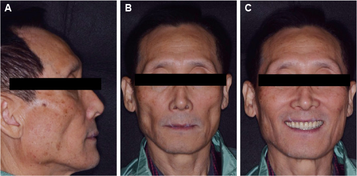
Fig. 9 Extraoral photograph after treatment. (A) Lateral view (B) Frontal view (C) Smile view.
Reference
-
1. Bartlett DW. The role of erosion in tooth wear: aetiology, prevention and management. Int Dent J. 2005; 55:277–284.
Article2. Smith BG, Bartlett DW, Robb ND. The prevalence, etiology and management of tooth wear in the United Kingdom. J Prosthet Dent. 1997; 78:367–372.
Article3. Gregory-Head B, Curtis DA. Erosion caused by gastroesophageal reflux: diagnostic considerations. J Prosthodont. 1997; 6:278–285.
Article4. Etman MK, Woolford M, Dunne S. Quantitative measurement of tooth and ceramic wear: in vivo study. Int J Prosthodont. 2008; 21:245–252.5. Lambrechts P, Braem M, Vuylsteke-Wauters M, Vanherle G. Quantitative in vivo wear of human enamel. J Dent Res. 1989; 68:1752–1754.
Article6. Verrett RG. Analyzing the etiology of an extremely worn dentition. J Prosthodont. 2001; 10:224–233.
Article7. Mulay G, Dugal R, Buhranpurwala M. An evaluation of wear of human enamel opposed by ceramics of different surface finishes. J Indian Prosthodont Soc. 2015; 15:111–118.
Article8. Johansson A, Haraldson T, Omar R, Kiliaridis S, Carlsson GE. A system for assessing the severity and progression of occlusal tooth wear. J Oral Rehabil. 1993; 20:125–131.
Article9. Mehta SB, Banerji S, Millar BJ, Suarez-Feito JM. Current concepts on the management of tooth wear: part 1. Assessment, treatment planning and strategies for the prevention and the passive management of tooth wear. Br Dent J. 2012; 212:17–27.
Article10. Briggs P, Bishop K. Fixed prostheses in the treatment of tooth wear. Eur J Prosthodont Restor Dent. 1997; 5:175–180.11. Sato S, Hotta TH, Pedrazzi V. Removable occlusal overlay splint in the management of tooth wear: a clinical report. J Prosthet Dent. 2000; 83:392–395.
Article12. Hemmings KW, Darbar UR, Vaughan S. Tooth wear treated with direct composite restorations at an increased vertical dimension: results at 30 months. J Prosthet Dent. 2000; 83:287–293.
Article13. Dahl BL, Krogstad O. The effect of a partial bite-raising splint on the inclination of upper and lower front teeth. Acta Odontol Scand. 1983; 41:311–314.
Article14. Ramfjord SP, Blankenship JR. Increased occlusal vertical dimension in adult monkeys. J Prosthet Dent. 1981; 45:74–83.
Article15. Turner KA, Missirlian DM. Restoration of the extremely worn dentition. J Prosthet Dent. 1984; 52:467–474.
Article16. Park JH, Jeong CM, Jeon YC, Lim JS. A study on the occlusal plane and the vertical dimension in Korean adults with natural dentition. J Korean Acad Prosthodont. 2005; 43:41–51.17. Jagger DC, Harrison A. An in vitro investigation into the wear effects of selected restorative materials on enamel. J Oral Rehabil. 1995; 22:275–281.
Article18. Lussi A. Dental erosion clinical diagnosis and case history taking. Eur J Oral Sci. 1996; 104:191–198.
Article19. Smith BG, Knight JK. An index for measuring the wear of teeth. Br Dent J. 1984; 156:435–438.
Article20. Wiley MG. Effects of porcelain on occluding surfaces of restored teeth. J Prosthet Dent. 1989; 61:133–137.
Article21. Jacobi R, Shillingburg HT Jr, Duncanson MG Jr. A comparison of the abrasiveness of six ceramic surfaces and gold. J Prosthet Dent. 1991; 66:303–309.
Article22. Dawson PE. Functional occlusion: from TMJ to smile design. Elsevier Health Sciences;2006.23. Rivera-Morales WC, Mohl ND. Relationship of occlusal vertical dimension to the health of the masticatory system. J Prosthet Dent. 1991; 65:547–553.
Article24. Dahl BL, Carlsson GE, Ekfeldt A. Occlusal wear of teeth and restorative materials. A review of classification, etiology, mechanisms of wear, and some aspects of restorative procedures. Acta Odontol Scand. 1993; 51:299–311.
Article25. Abduo J, Lyons K. Clinical considerations for increasing occlusal vertical dimension: a review. Aust Dent J. 2012; 57:2–10.
Article26. Aglietta M, Siciliano VI, Zwahlen M, Brägger U, Pjetursson BE, Lang NP, Salvi GE. A systematic review of the survival and complication rates of implant supported fixed dental prostheses with cantilever extensions after an observation period of at least 5 years. Clin Oral Implants Res. 2009; 20:441–451.
Article27. Zurdo J, Romão C, Wennström JL. Survival and complication rates of implant-supported fixed partial dentures with cantilevers: a systematic review. Clin Oral Implants Res. 2009; 20:59–66.
Article28. Pjetursson BE, Tan K, Lang NP, Brägger U, Egger M, Zwahlen M. A systematic review of the survival and complication rates of fixed partial dentures (FPDs) after an observation period of at least 5 years. Clin Oral Implants Res. 2004; 15:625–642.
Article29. Palmer RM, Howe LC, Palmer PJ, Wilson R. prospective clinical trial of single Astra Tech 4.0 or 5.0 diameter implants used to support two-unit cantilever bridges: results after 3 years. Clin Oral Implants Res. 2012; 23:35–40.
Article30. Wennström J, Zurdo J, Karlsson S, Ekestubbe A, Gröndahl K, Lindhe J. Bone level change at implant-supported fixed partial dentures with and without cantilever extension after 5 years in function. J Clin Periodontol. 2004; 31:1077–1083.
Article
- Full Text Links
-
- Actions
-
Cited
- CITED
-
- Close
- Share
-
- Similar articles
-
- Full mouth rehabilitation of the patient with severely worn dentition: a case report
- Full mouth rehabilitation in patient with loss of vertical dimension and deep bite due to tooth wear
- A case of full mouth rehabilitation with vertical dimension gaining in patient withseverely worn dentition and loss of vertical dimension due to loss of posterior support
- Full mouth rehabilitation with vertical dimension increase in patient with severely worn out dentition: A case report
- Full mouth rehabilitation in osteoporosis patient with loss of teeth and excessive wear
