The bactericidal effect of an atmospheric-pressure plasma jet on Porphyromonas gingivalis biofilms on sandblasted and acid-etched titanium discs
- Affiliations
-
- 1Department of Periodontology and Dental Research Institute, Seoul National University School of Dentistry, Seoul, Korea. yjseol@snu.ac.kr
- 2Program of Clinical Dental Education and Dental Research Institute, Seoul National University School of Dentistry, Seoul, Korea.
- 3Plasma Technology Research Center, National Fusion Research Institute, Gunsan, Korea.
- 4Department of Energy Systems (Nuclear) Engineering, Seoul National University School of Engineering, Seoul, Korea.
- KMID: 2461022
- DOI: http://doi.org/10.5051/jpis.2019.49.5.319
Abstract
- PURPOSE
Direct application of atmospheric-pressure plasma jets (APPJs) has been established as an effective method of microbial decontamination. This study aimed to investigate the bactericidal effect of direct application of an APPJ using helium gas (He-APPJ) on Porphyromonas gingivalis biofilms on sandblasted and acid-etched (SLA) titanium discs.
METHODS
On the SLA discs covered by P. gingivalis biofilms, an APPJ with helium (He) as a discharge gas was applied at 3 different time intervals (0, 3, and 5 minutes). To evaluate the effect of the plasma itself, the He gas-only group was used as the control group. The bactericidal effect of the He-APPJ was determined by the number of colony-forming units. Bacterial viability was observed by confocal laser scanning microscopy (CLSM), and bacterial morphology was examined by scanning electron microscopy (SEM).
RESULTS
As the plasma treatment time increased, the amount of P. gingivalis decreased, and the difference was statistically significant. In the SEM images, compared to the control group, the bacterial biofilm structure on SLA discs treated by the He-APPJ for more than 3 minutes was destroyed. In addition, the CLSM images showed consistent results. Even in sites distant from the area of direct He-APPJ exposure, decontamination effects were observed in both SEM and CLSM images.
CONCLUSIONS
He-APPJ application was effective in removing P. gingivalis biofilm on SLA titanium discs in an in vitro experiment.
Keyword
MeSH Terms
Figure
-
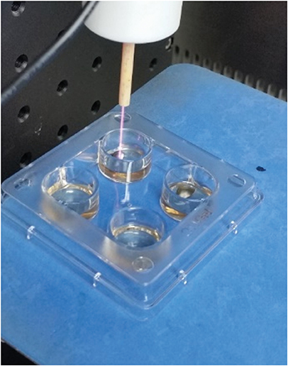
Figure 1 Photograph of the apparatus for He-APPJ generation and He-APPJ application to titanium discs in this study. He-APPJ: helium atmospheric-pressure plasma jet.
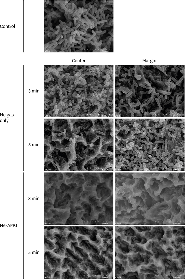
Figure 2 Scanning electron microscopy images of the central and marginal areas after He-APPJ treatment. The control group (no treatment) showed heavy bacterial accumulation on the discs. In the He gas–only treatment group, remaining bacterial biofilm was observed in both the central and marginal areas in the 3-minute treatment group, while bacterial rods had been eliminated from the central area of the 5-minute treatment group, but not in the marginal area. However, the He-APPJ group showed a clear titanium surface without a bacterial biofilm in both the 3-minute and 5-minute treatment groups. He-APPJ: helium atmospheric-pressure plasma jet.

Figure 3 Confocal laser scanning microscopy images of the central and marginal areas after He-APPJ application. Heavy live bacterial accumulation on the discs was observed in the control group. In the He gas–only treatment group, the amount of bacteria was higher in the central area than in the marginal area in the 3-minute treatment group. In the 5-minute treatment group, the amount of bacteria decreased in both the central and marginal areas, although more live bacteria were observed in the marginal area. in the He-APPJ treatment group, the amount of bacteria was lower in the central area than in the marginal area in both the 3-minute and 5-minute treatment groups. Only a few viable bacteria were observed in the central area of the 3-minute treatment group. In the marginal area, a mixture of live and dead bacteria was found, whereas most bacteria were dead in the 5-minute treatment group. He: helium, He-APPJ: helium atmospheric-pressure plasma jet.
Reference
-
1. Mombelli A. Microbiology and antimicrobial therapy of peri-implantitis. Periodontol 2000. 2002; 28:177–189.
Article2. Schwarz F, Sculean A, Romanos G, Herten M, Horn N, Scherbaum W, et al. Influence of different treatment approaches on the removal of early plaque biofilms and the viability of SAOS2 osteoblasts grown on titanium implants. Clin Oral Investig. 2005; 9:111–117.
Article3. Persson LG, Mouhyi J, Berglundh T, Sennerby L, Lindhe J. Carbon dioxide laser and hydrogen peroxide conditioning in the treatment of periimplantitis: an experimental study in the dog. Clin Implant Dent Relat Res. 2004; 6:230–238.
Article4. Schwarz F, Nuesry E, Bieling K, Herten M, Becker J. Influence of an erbium, chromium-doped yttrium, scandium, gallium, and garnet (Er,Cr:YSGG) laser on the reestablishment of the biocompatibility of contaminated titanium implant surfaces. J Periodontol. 2006; 77:1820–1827.
Article5. Schou S, Holmstrup P, Jørgensen T, Skovgaard LT, Stoltze K, Hjørting-Hansen E, et al. Implant surface preparation in the surgical treatment of experimental peri-implantitis with autogenous bone graft and ePTFE membrane in cynomolgus monkeys. Clin Oral Implants Res. 2003; 14:412–422.
Article6. Schwarz F, Ferrari D, Popovski K, Hartig B, Becker J. Influence of different air-abrasive powders on cell viability at biologically contaminated titanium dental implants surfaces. J Biomed Mater Res B Appl Biomater. 2009; 88:83–91.
Article7. Kreisler M, Kohnen W, Christoffers AB, Götz H, Jansen B, Duschner H, et al. In vitro evaluation of the biocompatibility of contaminated implant surfaces treated with an Er : YAG laser and an air powder system. Clin Oral Implants Res. 2005; 16:36–43.
Article8. Mendis DA, Rosenberg M, Azam F. A note on the possible electrostatic disruption of bacteria. IEEE Trans Plasma Sci. 2000; 28:1304–1306.
Article9. Laroussi M. Nonthermal decontamination of biological media by atmospheric-pressure plasmas: review, analysis, and prospects. IEEE Trans Plasma Sci. 2002; 30:1409–1415.
Article10. Kim JH, Lee MA, Han GJ, Cho BH. Plasma in dentistry: a review of basic concepts and applications in dentistry. Acta Odontol Scand. 2014; 72:1–12.
Article11. Lackmann JW, Bandow JE. Inactivation of microbes and macromolecules by atmospheric-pressure plasma jets. Appl Microbiol Biotechnol. 2014; 98:6205–6213.
Article12. Jablonowski L, Koban I, Berg MH, Kindel E, Duske K, Schröder K, et al. Elimination of E. Faecalis by a new non-thermal atmospheric pressure plasma handheld device for endodontic treatment. A preliminary investigation. Plasma Process Polym. 2013; 10:499–505.
Article13. Yang B, Chen J, Yu Q, Li H, Lin M, Mustapha A, et al. Oral bacterial deactivation using a low-temperature atmospheric argon plasma brush. J Dent. 2011; 39:48–56.
Article14. Cho BH, Han GJ, Oh KH, Chung SN, Chun BH. The effect of plasma polymer coating using atmospheric-pressure glow discharge on the shear bond strength of composite resin to ceramic. J Mater Sci. 2010; 46:2755–2763.
Article15. Nam SH, Lee HJ, Hong JW, Kim GC. Efficacy of nonthermal atmospheric pressure plasma for tooth bleaching. Sci World J. 2015; 2015:581731.
Article16. Canullo L, Peñarrocha D, Clementini M, Iannello G, Micarelli C. Impact of plasma of argon cleaning treatment on implant abutments in patients with a history of periodontal disease and thin biotype: radiographic results at 24-month follow-up of a RCT. Clin Oral Implants Res. 2015; 26:8–14.
Article17. Koban I, Holtfreter B, Hübner NO, Matthes R, Sietmann R, Kindel E, et al. Antimicrobial efficacy of non-thermal plasma in comparison to chlorhexidine against dental biofilms on titanium discs in vitro - proof of principle experiment. J Clin Periodontol. 2011; 38:956–965.
Article18. Carreiro AF, Delben JA, Guedes S, Silveira EJ, Janal MN, Vergani CE, et al. Low-temperature plasma on peri-implant-related biofilm and gingival tissue. J Periodontol. 2019; 90:507–515.
Article19. Yoon SY, Kim KH, Seol YJ, Kim SJ, Bae B, Huh SR, et al. Effects of metastable species in helium and argon atmospheric pressure plasma jets (APPJs) on inactivation of periodontopathogenic bacteria. J Korean Phys Soc. 2016; 68:1176–1191.
Article20. Berglundh T, Gotfredsen K, Zitzmann NU, Lang NP, Lindhe J. Spontaneous progression of ligature induced peri-implantitis at implants with different surface roughness: an experimental study in dogs. Clin Oral Implants Res. 2007; 18:655–661.
Article21. Ehlbeck J, Schnabel U, Polak M, Winter J, von Woedtke T, Brandenburg R, et al. Low temperature atmospheric pressure plasma sources for microbial decontamination. J Phys D Appl Phys. 2010; 44:013002.
Article22. Laroussi M, Mendis DA, Rosenberg M. Plasma interaction with microbes. New J Phys. 2003; 5:41.1–41.10.
Article23. Venezia RA, Orrico M, Houston E, Yin SM, Naumova YY. Lethal activity of nonthermal plasma sterilization against microorganisms. Infect Control Hosp Epidemiol. 2008; 29:430–436.
Article24. Laroussi M, Sayler GS, Glascock BB, McCurdy B, Pearce ME, Bright NG, et al. Images of biological samples undergoing sterilization by a glow discharge at atmospheric pressure. IEEE Trans Plasma Sci. 1999; 27:34–35.
Article25. Lee K, Paek KH, Ju WT, Lee Y. Sterilization of bacteria, yeast, and bacterial endospores by atmospheric-pressure cold plasma using helium and oxygen. J Microbiol. 2006; 44:269–275.26. Montie TC, Kelly-Wintenberg K, Roth JR. An overview of research using the one atmosphere uniform glow discharge plasma (OAUGDP) for sterilization of surfaces and materials. IEEE Trans Plasma Sci. 2000; 28:41–50.
Article27. Kim SJ, Yoon SY, Kim GH. Bullet velocity distribution of a helium atmospheric-pressure plasma jet in various N2/O2 mixed ambient conditions. IEEE Trans Plasma Sci. 2015; 43:2054–2063.
Article28. Miura T, Egawa M, Ito T, Eguro T, Tanabe K, Yoshinari M. Debridement effect on periodontal pathogen Porphyromonas gingivalis cultured on titanium by application of atmospheric-pressure plasma. J Biomed Sci Eng. 2017; 10:51–59.
Article29. Kim MS, Lee JS, Shin HK, Kim JS, Yun JH, Cho KS. Prospective randomized, controlled trial of sinus grafting using Escherichia-coli-produced rhBMP-2 with a biphasic calcium phosphate carrier compared to deproteinized bovine bone. Clin Oral Implants Res. 2015; 26:1361–1368.
Article30. Lamont RJ, Jenkinson HF. Life below the gum line: pathogenic mechanisms of Porphyromonas gingivalis . Microbiol Mol Biol Rev. 1998; 62:1244–1263.
Article31. Mahasneh A, Darby M, Tolle SL, Hynes W, Laroussi M, Karakas E. Inactivation of Porphyromonas gingivalis by low-temperature atmospheric pressure plasma. Plasma Med. 2011; 1:191–204.
Article32. Marsh PD. Dental plaque as a microbial biofilm. Caries Res. 2004; 38:204–211.
Article33. Preissner S, Wirtz HC, Tietz AK, Abu-Sirhan S, Herbst SR, Hartwig S, et al. Bactericidal efficacy of tissue tolerable plasma on microrough titanium dental implants: An in-vitro-study. J Biophotonics. 2016; 9:637–644.
Article34. Sladek RE, Filoche SK, Sissons CH, Stoffels E. Treatment of Streptococcus mutans biofilms with a nonthermal atmospheric plasma. Lett Appl Microbiol. 2007; 45:318–323.
Article35. Berney M, Hammes F, Bosshard F, Weilenmann HU, Egli T. Assessment and interpretation of bacterial viability by using the LIVE/DEAD BacLight Kit in combination with flow cytometry. Appl Environ Microbiol. 2007; 73:3283–3290.
Article36. Shashurin A, Keidar M, Bronnikov S, Jurjus RA, Stepp MA. Living tissue under treatment of cold plasma atmospheric jet. Appl Phys Lett. 2008; 93:181501.
Article37. Vleugels M, Shama G, Deng XT, Greenacre E, Brocklehurst T, Kong MG. Atmospheric plasma inactivation of biofilm-forming bacteria for food safety control. IEEE Trans Plasma Sci. 2005; 33:824–828.
Article38. Fridman G, Brooks AD, Balasubramanian M, Fridman A, Gutsol A, Vasilets VN, et al. Comparison of direct and indirect effects of non-thermal atmospheric-pressure plasma on bacteria. Plasma Process Polym. 2007; 4:370–375.
Article
- Full Text Links
-
- Actions
-
Cited
- CITED
-
- Close
- Share
-
- Similar articles
-
- Efficacy of an LED toothbrush on a Porphyromonas gingivalis biofilm on a sandblasted and acid-etched titanium surface: an in vitro study
- Environmental factors regulating the expression of Porphyromonas gingivalis heat shock protein
- Fusobacterium nucleatum modulates serum binding to Porphyromonas gingivalis biofilm
- The relation between metabolic acid and the virulence of porphyromonas gingivalis serotype b
- A comparative clinical study on oxidized titanium implants and sandblasted large-grit acid etched implants in soft bone
