J Korean Acad Periodontol.
2009 Aug;39(Suppl):205-212.
A comparative clinical study on oxidized titanium implants and sandblasted large-grit acid etched implants in soft bone
- Affiliations
-
- 1Department of Periodontology, Research Institute for Periodontal Regeneration, College of Dentistry, Yonsei University, Korea. kscho@yuhs.ac
Abstract
-
PURPOSE: The aim of this retrospective study was to compare the survival rate of oxidized titanium implants and sandblasted large-grit acid etched implants in soft bone.
METHODS
201 oxidized titanium implants were inserted in 84 patients between May 1999 and May 2004. 120 sandblasted large-grit acid etched implants were inserted in 74 patients between December 2000 and May 2004. The patients were followed-up 0~5 years in ITI group or 0~6 years in BRA group, respectively. The following information was collected from the patient records: age, gender, systemic disease, implant type, number, length and diameter of the implants, their location in the jaws, bone quantity, the number of failed implants, the causes of failure, and advanced surgery for bone augmentation.
RESULTS
In the oxidized titanium implants, 8 implants showed early failure, and 1 implant showed late failure, respectively. The cumulative survival rate was 95.48%. In the sandblasted large-grit acid etched implants, 1 implant showed late failure and cumulative survival rate was 99.10%. The cumulative survival rate and the survival rates in the case of the advanced procedure during the implant placement were not significantly different in both groups.
CONCLUSIONS
Oxidized titanium implants and sandblasted large-grit acid etched implants can be used successfully in soft bone regardless of the surgical methods used during the implant placement.
Keyword
MeSH Terms
Figure
-
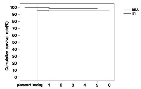
Figure 1 Cumulative survival rates in relation to the implant type (BRA: Brånemark Ti-Unite™ implants, ITI: ITI SLA implants).
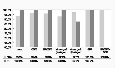
Figure 2 Implant survival rate according to the additional surgical procedures and implant type (BRA: Brånemark Ti-Unite™ implants, ITI: ITI SLA implants).
Reference
-
1. Albrektsson T, Brånemark PI, Hansson HA, Lindstrom J. Titanium implants. Requirements for ensuring a long-lasting direct bone anchorage in man. Acta Orthop Scand. 1981. 52:155–170.
Article2. Martinez H, Davarpanah M, Missika P, Celletti R, Lazzara R. Optimal implant stabilization in low density bone. Clin Oral Implants Res. 2001. 12:423–432.
Article3. Boyne PJ, James RA. Grafting of the maxillary sinus floor with autogenous marrow and bone. J Oral Surg. 1980. 38:613–616.4. Tatum OH. Maxillary and sinus implant reconstruction. Dent Clin North Am. 1986. 30:207–229.5. Summers RB. The osteotome technique: Part III: Less invasive methods of elevating the sinus floor. Compendium. 1994. 15:698700702–704.6. Buser D, Dula K, Lang NP, Nyman S. Long-term stability of osseointegrated implants in bone regenerated with the membrane technique. Five-year results of a prostective study with 12 implants. Clin Oral Implants Res. 1996. 7:175–183.
Article7. Fugazzotto PA. Success and failure rates of osseointegrated implants in function in regenerated bone for 72 to 133 months. Int J Oral Maxillofac Implants. 2005. 20:77–83.8. Summers RB. A new concept in maxillary implant surgery: The osteotome technique. Compendium. 1994. 15:152154–156. 1589. Pinholt EM. Brånemark and ITI dental implants in the human bone grafted maxilla: a comparative evaluation. Clin Oral Implants Res. 2003. 14:584–592.
Article10. Åstrand P, Engquist B, Anzén B, et al. A three-year follow-up report of a comparative study of ITI dental implants® and Brånemark System® implants in the treatment of the partially edentulous maxilla. Clin Implant Dent Relat Res. 2004. 6:130–141.
Article11. Kang NW, Jung UW, Choi SH, et al. Bone added osteotome sinus floor elevation with simultaneous placement of Brånamark Ti-Unite and ITI SLA implants. J Korean Acad Periodontol. 2005. 35:609–621.
Article12. Hong SB, Chai GJ, Jung UW, et al. Clinical evaluation of Brånemark Ti-Unite implant and ITI SLA implant in the post maxillary area with sinus elevation technique. J Korean Acad Periodontol. 2005. 35:813–822.
Article13. Lekholm U, Zarb GA. Brånemark PI, Zarb GA, Albretksson T, editors. Patient selection and preparations. Tissue-integrated prostheses: Osseointegration in clinical dentistry. 1985. 1st ed. Chicago: Quintessence Publishing Co Inc;199–220.14. Buser D, Weber HP, Lang NP. Tissue integration of non-submerged implants: 1-year results of a prospective study with 100 ITI hollow-cylinder and hollow-screw implants. Clin Oral Implants Res. 1990. 1:33–40.
Article15. Cutler SJ, Ederer F. Maximum utilization of the life table method in analyzing survival. J Chronic Dis. 1958. 6:699–712.
Article16. Engquist B, Bergendal T, Kallus T, Linden U. A retrospective multicenter evaluation of osseointegrated implants supporting overdentures. Int J Oral Maxillofac Implants. 1988. 3:129–134.17. Friberg B, Jemt T, Lekholm U. Early failures in 4,641 consecutively placed Brånemark dental implants: a study from stage I surgery to the connection of completed prostheses. Int J Oral Maxillofac Implants. 1988. 3:129–134.18. Jaffin R, Berman C. The excessive loss of Brånemark fixtures in type IV bone: a 5-year analysis. J Periodontol. 1991. 62:2–4.
Article19. Wilke HJ, Claes L, Stenemann S. Heimke G, Soltesz U, Lee AJC, editors. Clinical implant material. Advances in biomaterials. 1990. Vol. 9. Amsterdam: Elsevier Science Publishing Co Inc;390.20. Buser D, Nydegger T, Hirt HP, Cochran DL, Nolte LP. Removal torque values of titanium implants in the maxilla of miniature pigs. Int J Oral Maxillofac Implants. 1998. 13:611–619.21. Henry P, Tan A, Allan B, Hall J, Johansson C. Removal torque comparison of TiUnite and turned implants in the Greyhound dog mandible. Appl Osseointegration Res. 2000. 1:15–17.22. Glauser R, Portmann M, Ruhstaller P, et al. Stability measurements of immediately loaded machined and oxidized implants in the posterior maxilla. A comparative clinical study using resonance frequency analysis. Appl Osseointegration Res. 2001. 2:27–29.23. Lee ES, Ahn YB, Lee WJ, Kim HS. Survival rate of implant placement in the maxilla treated with sinus elevation by the lateral approach: A retrospective study. J Korean Acad Periodontol. 2008. 38:589–594.
Article24. Glauser R, Ruhstaller P, Windisch S, et al. Immediate occlusal loading of Brånemark TiUnite™ implants placed predominantly in soft bone: 4-year results of a prospective clinical study. Clin Implant Dent Relat Res. 2005. 7:s52–s59.25. Friberg B, Dahlin C, Widmark G, Östman PO, Billström C. One-year results of a prospective multicenter study on Brånemark System® implants with a TiUnite™ surface. Clin Implant Dent Relat Res. 2005. 7:s70–s75.26. Stricker A, Voss PJ, Gutwald R, Schramm A, Schmelzeisen R. Maxillary sinus floor augmentation with autogenous bone grafts to enable placement of SLA-surfaced implants: preliminary results after 15-40 months. Clin Oral Implants Res. 2003. 14:207–212.
Article27. Glauser R, Gottlow J, Lundgren AK, et al. Immediate occlusal loading of Brånemark Mk IV TiUnite™ implants placed in bone quality type 4. Appl Osseointegration Res. 2002. 3:22–24.28. Vanden Bogaerde L, Pedretti G, Dellacasa P, et al. Early function of splinted implants in maxillas and posterior mandibles, using Brånemark System® TiUnite™ implants: An 18-month prospective clinical multicenter study. Clin Implant Dent Relat Res. 2004. 6:121–129.
Article29. Rocci A, Martignoni M, Gottlow J, Rangert B. Immediate function of single and partial reconstructions in the maxilla using Mk IV fixtures: A retrospective analysis. Appl Osseointegration Res. 2001. 2:22–26.30. Bornstein MM, Schmid B, Belser UC, Lussi A, Buser D. Early loading of nonsubmerged titanium implants with a sandblasted and acid-etched (SLA) surface: 5-year results of a prospective study in partially edentulous patients. Clin Oral Implants Res. 2005. 16:631–638.
Article31. Ferrigno N, Laureti M. Surgical advantages with ITI TE® implants placement in conjunction with split crest technique: 18-month results of an ongoing prospective study. Clin Oral Implants Res. 2005. 16:147–155.
Article32. Fugazzotto PA, Vlassis J, Butler B. ITI implant use in private practice: Clinical results with 5,526 implants followed up to 72+ months in function. Int J Oral Maxillofac Implants. 2004. 19:408–412.33. Luongo G, Raimondo R, Filippini P, Gualini F, Paoleschi C. Early loading of sandblasted, acid-etched implants in the posterior maxilla and mandible: A 1-year follow-up report from a multicenter 3-year prospective study. Int J Oral Maxillofac Implants. 2005. 20:84–91.34. Nedir R, Bischof M, Briaux JM, et al. A 7-year life table analysis from a prospective study on ITI implants with special emphasis on the use of short implants: Results from a private practice. Clin Oral Implants Res. 2004. 15:150–157.
Article35. Nordin T, Nilsson R, Frykholm A, Hallman M. A 3-arm study of early loading of rough-surfaced implants in the completely edentulous maxilla and in the edentulous posterior maxilla and mandible: results after 1 year of loading. Int J Oral Maxillofac Implants. 2004. 19:880–886.36. Salvi GE, Gallini G, Lang NP. Early loading (2 or 6 weeks) of sandblasted and acid-etched (SLA) ITI® implants in the posterior mandible: A 1-year randomized controlled clinical trial. Clin Oral Implants Res. 2004. 15:142–149.
Article37. Vanden Bogaerde L, Rangert B, Wendelhag I. Immediate / early function of Brånemark System® TiUnite™ implants in fresh extraction sockets in maxillae and posterior mandibles: An 18-month prospective clinical study. Clin Implant Dent Relat Res. 2005. 7:s121–s130.
- Full Text Links
-
- Actions
-
Cited
- CITED
-
- Close
- Share
-
- Similar articles
-
- Scanning Electron Microscopic Study of the Effects of Citric Acid on the Change of Implant Surface According to Application Time
- Characteristics of contact and distance osteogenesis around modified implant surfaces in rabbit tibiae
- Histomorphometric and Removal Torque Values Comparision of Rough Surface Titanium Implants
- The Effect of N-acetyl cysteine (NAC) loading on the bone formation surrounding sandblastedand large-grit and acid-etched implants in the dog: A pilot study
- Retrospective clinical study of an implant with a sandblasted, large-grit, acid-etched surface and internal connection: analysis of short-term success rate and marginal bone loss
