Triple Arterial Phase MR Imaging with Gadoxetic Acid Using a Combination of Contrast Enhanced Time Robust Angiography, Keyhole, and Viewsharing Techniques and Two-Dimensional Parallel Imaging in Comparison with Conventional Single Arterial Phase
- Affiliations
-
- 1Department of Radiology, Seoul National University Hospital, Seoul 03080, Korea. jmlshy2000@gmail.com
- 2Department of Radiology, Seoul National University College of Medicine, Seoul 03087, Korea.
- 3Institute of Radiation Medicine, Seoul National University Medical Research Center, Seoul 03087, Korea.
- 4Department of Radiology, Konkuk University Medical Center, Seoul 05030, Korea.
- 5Philips Healthcare Korea, Seoul 04342, Korea.
- KMID: 2455422
- DOI: http://doi.org/10.3348/kjr.2016.17.4.522
Abstract
OBJECTIVE
To determine whether triple arterial phase acquisition via a combination of Contrast Enhanced Time Robust Angiography, keyhole, temporal viewsharing and parallel imaging can improve arterial phase acquisition with higher spatial resolution than single arterial phase gadoxetic-acid enhanced magnetic resonance imaging (MRI).
MATERIALS AND METHODS
Informed consent was waived for this retrospective study by our Institutional Review Board. In 752 consecutive patients who underwent gadoxetic acid-enhanced liver MRI, either single (n = 587) or triple (n = 165) arterial phases was obtained in a single breath-hold under MR fluoroscopy guidance. Arterial phase timing was assessed, and the degree of motion was rated on a four-point scale. The percentage of patients achieving the late arterial phase without significant motion was compared between the two methods using the χ2 test.
RESULTS
The late arterial phase was captured at least once in 96.4% (159/165) of the triple arterial phase group and in 84.2% (494/587) of the single arterial phase group (p < 0.001). Significant motion artifacts (score ≤ 2) were observed in 13.3% (22/165), 1.2% (2/165), 4.8% (8/165) on 1st, 2nd, and 3rd scans of triple arterial phase acquisitions and 6.0% (35/587) of single phase acquisitions. Thus, the late arterial phase without significant motion artifacts was captured in 96.4% (159/165) of the triple arterial phase group and in 79.9% (469/587) of the single arterial phase group (p < 0.001).
CONCLUSION
Triple arterial phase imaging may reliably provide adequate arterial phase imaging for gadoxetic acid-enhanced liver MRI.
Keyword
MeSH Terms
-
Adult
Aged
Aged, 80 and over
Angiography
Arteries/diagnostic imaging/pathology
Artifacts
Contrast Media/*chemistry
Female
Fluoroscopy
Gadolinium DTPA/*chemistry
Humans
Image Interpretation, Computer-Assisted
Liver Neoplasms/diagnosis/diagnostic imaging/pathology
*Magnetic Resonance Imaging
Male
Middle Aged
Neoplasms/*diagnosis/diagnostic imaging/pathology
Retrospective Studies
Young Adult
Contrast Media
Gadolinium DTPA
Figure
-
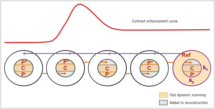
Fig. 1 K-space acquisition scheme using combination of CENTRA, keyhole and viewsharing.K-space is divided into central (Kz-Ky) and peripheral portion (R), and central K-space is sampled repeatedly in random manner whilst peripheral K-space is acquired once last to serve as reference scan for each reconstruction (violet arrows). Central K-space is divided into three keyhole fractions including central (C) and two peripheries (P+, P-) and fractions are acquired in alternating fashion between combinations of (P+, C) and (C, P-). To avoid signal discontinuation, P+ or P- of keyhole obtained on previous scan is added to reconstruction of next scan (orange arrows). CENTRA = contrast enhanced time robust angiography
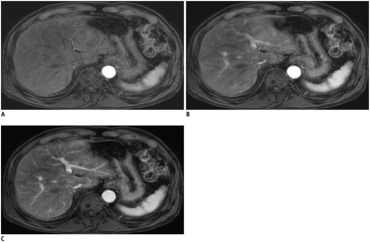
Fig. 2 Triple arterial phase (TAP) of 80-year-old man with colon cancer.1st, 2nd, and 3rd TAP scans (A-C) provide early arterial phase (A) and two late arterial phases (B, C) with different parenchymal enhancement degrees.
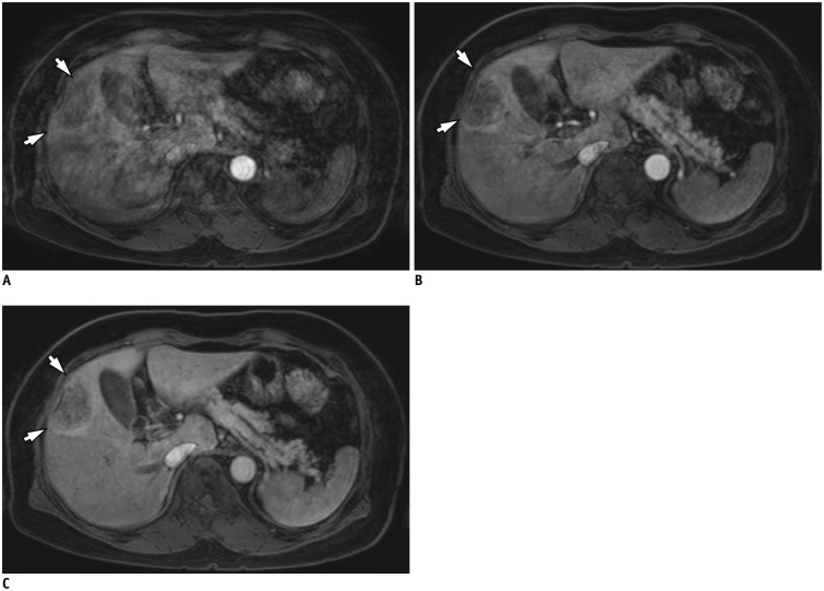
Fig. 3 Fig. 3. Triple arterial phase of 70-year-old woman with colon cancer liver metastasis.1st scan was deteriorated by significant motion artifacts with truncation artifact, leading to non-diagnostic image (A). Following 2nd (B) and 3rd (C) TAP scans were regarded as optimal late arterial phase imaging, and successfully demonstrate 4 cm rim enhancing mass which was confirmed as metastasis after surgery (arrows). TAP = triple arterial phase
Cited by 3 articles
-
The Diagnostic Performance of Liver MRI without Intravenous Contrast for Detecting Hepatocellular Carcinoma: A Case-Controlled Feasibility Study
Seunghee Han, Joon-Il Choi, Michael Yong Park, Moon Hyung Choi, Sung Eun Rha, Young Joon Lee
Korean J Radiol. 2018;19(4):568-577. doi: 10.3348/kjr.2018.19.4.568.Rapid Imaging: Recent Advances in Abdominal MRI for Reducing Acquisition Time and Its Clinical Applications
Jeong Hee Yoon, Marcel Dominik Nickel, Johannes M. Peeters, Jeong Min Lee
Korean J Radiol. 2019;20(12):1597-1615. doi: 10.3348/kjr.2018.0931.Advanced Methods in Dynamic Contrast Enhanced Arterial Phase Imaging of the Liver
Yoon-Chul Kim
Investig Magn Reson Imaging. 2019;23(1):1-16. doi: 10.13104/imri.2019.23.1.1.
Reference
-
1. Choi JY, Lee JM, Sirlin CB. CT and MR imaging diagnosis and staging of hepatocellular carcinoma: part II. Extracellular agents, hepatobiliary agents, and ancillary imaging features. Radiology. 2014; 273:30–50. PMID: 25247563.
Article2. Sun HY, Lee JM, Shin CI, Lee DH, Moon SK, Kim KW, et al. Gadoxetic acid-enhanced magnetic resonance imaging for differentiating small hepatocellular carcinomas (< or =2 cm in diameter) from arterial enhancing pseudolesions: special emphasis on hepatobiliary phase imaging. Invest Radiol. 2010; 45:96–103. PMID: 20057319.3. Lee YJ, Lee JM, Lee JS, Lee HY, Park BH, Kim YH, et al. Hepatocellular carcinoma: diagnostic performance of multidetector CT and MR imaging-a systematic review and meta-analysis. Radiology. 2015; 275:97–109. PMID: 25559230.
Article4. Kudo M, Matsui O, Izumi N, Iijima H, Kadoya M, Imai Y. Liver Cancer Study Group of Japan. Surveillance and diagnostic algorithm for hepatocellular carcinoma proposed by the Liver Cancer Study Group of Japan: 2014 update. Oncology. 2014; 87(Suppl 1):7–21. PMID: 25427729.
Article5. Mitchell DG, Bruix J, Sherman M, Sirlin CB. LI-RADS (Liver Imaging Reporting and Data System): summary, discussion, and consensus of the LI-RADS Management Working Group and future directions. Hepatology. 2015; 61:1056–1065. PMID: 25041904.
Article6. Hope TA, Fowler KJ, Sirlin CB, Costa EA, Yee J, Yeh BM, et al. Hepatobiliary agents and their role in LI-RADS. Abdom Imaging. 2015; 40:613–625. PMID: 25287679.
Article7. Korean Liver Cancer Study Group (KLCSG). National Cancer Center, Korea (NCC). 2014 Korean Liver Cancer Study Group-National Cancer Center Korea practice guideline for the management of hepatocellular carcinoma. Korean J Radiol. 2015; 16:465–522. PMID: 25995680.8. Zech CJ, Vos B, Nordell A, Urich M, Blomqvist L, Breuer J, et al. Vascular enhancement in early dynamic liver MR imaging in an animal model: comparison of two injection regimen and two different doses Gd-EOB-DTPA (gadoxetic acid) with standard Gd-DTPA. Invest Radiol. 2009; 44:305–310. PMID: 19462484.
Article9. Pietryga JA, Burke LM, Marin D, Jaffe TA, Bashir MR. Respiratory motion artifact affecting hepatic arterial phase imaging with gadoxetate disodium: examination recovery with a multiple arterial phase acquisition. Radiology. 2014; 271:426–434. PMID: 24475864.
Article10. Davenport MS, Caoili EM, Kaza RK, Hussain HK. Matched within-patient cohort study of transient arterial phase respiratory motion-related artifact in MR imaging of the liver: gadoxetate disodium versus gadobenate dimeglumine. Radiology. 2014; 272:123–131. PMID: 24617733.
Article11. Tamada T, Ito K, Yoshida K, Kanki A, Higaki A, Tanimoto D, et al. Comparison of three different injection methods for arterial phase of Gd-EOB-DTPA enhanced MR imaging of the liver. Eur J Radiol. 2011; 80:e284–e288. PMID: 21296514.
Article12. Kim SM, Heo SH, Kim JW, Lim HS, Shin SS, Jeong YY, et al. Hepatic arterial phase on gadoxetic acid-enhanced liver MR imaging: a randomized comparison of 0.5 mL/s and 1 mL/s injection rates. Korean J Radiol. 2014; 15:605–612. PMID: 25246821.
Article13. Motosugi U, Ichikawa T, Sano K, Sou H, Onohara K, Muhi A, et al. Double-dose gadoxetic acid-enhanced magnetic resonance imaging in patients with chronic liver disease. Invest Radiol. 2011; 46:141–145. PMID: 21139506.
Article14. Park YS, Lee CH, Kim IS, Kiefer B, Woo ST, Kim KA, et al. Usefulness of controlled aliasing in parallel imaging results in higher acceleration in gadoxetic acid-enhanced liver magnetic resonance imaging to clarify the hepatic arterial phase. Invest Radiol. 2014; 49:183–188. PMID: 24276676.
Article15. Davenport MS, Bashir MR, Pietryga JA, Weber JT, Khalatbari S, Hussain HK. Dose-toxicity relationship of gadoxetate disodium and transient severe respiratory motion artifact. AJR Am J Roentgenol. 2014; 203:796–802. PMID: 25055154.
Article16. Hope TA, Saranathan M, Petkovska I, Hargreaves BA, Herfkens RJ, Vasanawala SS. Improvement of gadoxetate arterial phase capture with a high spatio-temporal resolution multiphase three-dimensional SPGR-Dixon sequence. J Magn Reson Imaging. 2013; 38:938–945. PMID: 23371926.
Article17. Beck GM, De Becker J, Jones AC, von Falkenhausen M, Willinek WA, Gieseke J. Contrast-enhanced timing robust acquisition order with a preparation of the longitudinal signal component (CENTRA plus) for 3D contrast-enhanced abdominal imaging. J Magn Reson Imaging. 2008; 27:1461–1467. PMID: 18504734.
Article18. Hadizadeh DR, Gieseke J, Beck G, Geerts L, Kukuk GM, Boström A, et al. View-sharing in keyhole imaging: partially compressed central k-space acquisition in time-resolved MRA at 3.0 T. Eur J Radiol. 2011; 80:400–406. PMID: 20447790.19. Saranathan M, Rettmann DW, Hargreaves BA, Clarke SE, Vasanawala SS. DIfferential Subsampling with Cartesian Ordering (DISCO): a high spatio-temporal resolution Dixon imaging sequence for multiphasic contrast enhanced abdominal imaging. J Magn Reson Imaging. 2012; 35:1484–1492. PMID: 22334505.
Article20. Fujinaga Y, Ohya A, Tokoro H, Yamada A, Ueda K, Ueda H, et al. Radial volumetric imaging breath-hold examination (VIBE) with k-space weighted image contrast (KWIC) for dynamic gadoxetic acid (Gd-EOB-DTPA)-enhanced MRI of the liver:advantages over Cartesian VIBE in the arterial phase. Eur Radiol. 2014; 24:1290–1299. PMID: 24633374.21. Agrawal MD, Spincemaille P, Mennitt KW, Xu B, Wang Y, Dutruel SP, et al. Improved hepatic arterial phase MRI with 3-second temporal resolution. J Magn Reson Imaging. 2013; 37:1129–1136. PMID: 23197440.
Article22. Kim KW, Lee JM, Jeon YS, Kang SE, Baek JH, Han JK, et al. Free-breathing dynamic contrast-enhanced MRI of the abdomen and chest using a radial gradient echo sequence with K-space weighted image contrast (KWIC). Eur Radiol. 2013; 23:1352–1360. PMID: 23187728.
Article23. Yoon JH, Lee JM, Yu MH, Kim EJ, Han JK, Choi BI. High-resolution T1-weighted gradient echo imaging for liver MRI using parallel imaging at high-acceleration factors. Abdom Imaging. 2014; 39:711–721. PMID: 24557640.
Article24. Yu MH, Lee JM, Yoon JH, Kiefer B, Han JK, Choi BI. Clinical application of controlled aliasing in parallel imaging results in a higher acceleration (CAIPIRINHA)-volumetric interpolated breathhold (VIBE) sequence for gadoxetic acid-enhanced liver MR imaging. J Magn Reson Imaging. 2013; 38:1020–1026. PMID: 23559147.
Article25. Budjan J, Ong M, Riffel P, Morelli JN, Michaely HJ, Schoenberg SO, et al. CAIPIRINHA-Dixon-TWIST (CDT)-volumeinterpolated breath-hold examination (VIBE) for dynamic liver imaging: comparison of gadoterate meglumine, gadobutrol and gadoxetic acid. Eur J Radiol. 2014; 83:2007–2012. PMID: 25172427.
Article26. Merkle EM, Dale BM. Abdominal MRI at 3.0 T: the basics revisited. AJR Am J Roentgenol. 2006; 186:1524–1532. PMID: 16714640.
Article27. Willinek WA, Gieseke J, Conrad R, Strunk H, Hoogeveen R, von Falkenhausen M, et al. Randomly segmented central k-space ordering in high-spatial-resolution contrast-enhanced MR angiography of the supraaortic arteries: initial experience. Radiology. 2002; 225:583–588. PMID: 12409598.
Article28. Yoon JH, Lee JM, Yu MH, Kim EJ, Han JK, Choi BI. Fat-suppressed, three-dimensional T1-weighted imaging using high-acceleration parallel acquisition and a dual-echo Dixon technique for gadoxetic acid-enhanced liver MRI at 3 T. Acta Radiol. 2015; 56:1454–1462. PMID: 25480475.29. Hoogeveen R, von Falkenhausen M, Gieseke J. Fast dynamic, high resolution contrast-enhanced MR angiography with CENTRA keyhole and SENSE [abstract]. Proc Int Soc Mag Reson Med. 2004; 12:9.30. Tanimoto A, Higuchi N, Ueno A. Reduction of ringing artifacts in the arterial phase of gadoxetic acid-enhanced dynamic MR imaging. Magn Reson Med Sci. 2012; 11:91–97. PMID: 22790295.
Article31. Huh J, Kim SY, Yeh BM, Lee SS, Kim KW, Wu EH, et al. Troubleshooting arterial-phase MR images of gadoxetate disodium-enhanced liver. Korean J Radiol. 2015; 16:1207–1215. PMID: 26576109.
Article32. Landis JR, Koch GG. An application of hierarchical kappa-type statistics in the assessment of majority agreement among multiple observers. Biometrics. 1977; 33:363–374. PMID: 884196.
Article33. Haradome H, Grazioli L, Tsunoo M, Tinti R, Frittoli B, Gambarini S, et al. Can MR fluoroscopic triggering technique and slow rate injection provide appropriate arterial phase images with reducing artifacts on gadoxetic acid-DTPA (Gd-EOB-DTPA)-enhanced hepatic MR imaging? J Magn Reson Imaging. 2010; 32:334–340. PMID: 20677259.
Article34. Sharma P, Kalb B, Kitajima HD, Salman KN, Burrow B, Ray GL, et al. Optimization of single injection liver arterial phase gadolinium enhanced MRI using bolus track real-time imaging. J Magn Reson Imaging. 2011; 33:110–118. PMID: 21182128.
Article35. Nakamura S, Nakaura T, Kidoh M, Utsunomiya D, Doi Y, Harada K, et al. Timing of the hepatic arterial phase at Gd-EOBDTPA-enhanced hepatic dynamic MRI: comparison of the testinjection and the fixed-time delay method. J Magn Reson Imaging. 2013; 38:548–554. PMID: 23744782.
Article36. Hussain HK, Londy FJ, Francis IR, Nghiem HV, Weadock WJ, Gebremariam A, et al. Hepatic arterial phase MR imaging with automated bolus-detection three-dimensional fast gradientrecalled-echo sequence: comparison with test-bolus method. Radiology. 2003; 226:558–566. PMID: 12563155.
Article37. Cho JY, Lee YJ, Han HS, Yoon YS, Kim J, Choi Y, et al. Role of gadoxetic acid-enhanced magnetic resonance imaging in the preoperative evaluation of small hepatic lesions in patients with colorectal cancer. World J Surg. 2015; 39:1161–1166. PMID: 25609116.
Article38. Yoon JH, Lee JM, Yu MH, Kim EJ, Han JK, Choi BI. Fatsuppressed, three-dimensional T1-weighted imaging using high-acceleration parallel acquisition and a dual-echo Dixon technique for gadoxetic acid-enhanced liver MRI at 3 T. Acta Radiol. 2015; 56:1454–1462. PMID: 25480475.39. AlObaidy M, Ramalho M, Busireddy KK, Liu B, Burke LM, Altun E, et al. High-resolution 3D-GRE imaging of the abdomen using controlled aliasing acceleration technique - a feasibility study. Eur Radiol. 2015; 25:3596–3605. PMID: 25916391.
Article40. Wile GE, Leyendecker JR. Magnetic resonance imaging of the liver: sequence optimization and artifacts. Magn Reson Imaging Clin N Am. 2010; 18:525–547. PMID: 21094454.
Article
- Full Text Links
-
- Actions
-
Cited
- CITED
-
- Close
- Share
-
- Similar articles
-
- Advanced Methods in Dynamic Contrast Enhanced Arterial Phase Imaging of the Liver
- Triple Arterial Phase Hepatic MRI Using Four Dimensional T1-Weighted High Resolutions Imaging with Volume Excitation Keyhole Techniques: Feasibility and Initial Clinical Experience in Focal Liver Lesions
- Current Limitations and Potential Breakthroughs for the Early Diagnosis of Hepatocellular Carcinoma
- Hepatic Angiomyolipoma Presenting as a Hyperintense Lesion During the Hepatobiliary Phase of Gadoxetic Acid Enhanced-MRI: a Case Report
- Hepatic Arterial Phase on Gadoxetic Acid-Enhanced Liver MR Imaging: A Randomized Comparison of 0.5 mL/s and 1 mL/s Injection Rates
