Full mouth rehabilitation of a patient with severe tooth erosion with a digital crown lengthening guide
- Affiliations
-
- 1Department of Prosthodontics, School of Dentistry, Kyung Hee University, Seoul, Republic of Korea. odontopia@khu.ac.kr
- KMID: 2453783
- DOI: http://doi.org/10.4047/jkap.2019.57.3.280
Abstract
- In the oral cavity, the teeth undergo wear and corrosion throughout their lives. Progressive and constant tooth wear is a natural phenomenon of aging, but wear and corrosion due to specific factors are pathological factors. It can cause pathological damage of the occlusal surface, aesthetic problems, dimensional loss and jaw joint disorders. This case is a 26-year-old female patient with general tooth abrasion and erosion on the entire dentition. Diagnostic wax-up was fabricated based on the information including digital facial analysis, physiological stabilization, and evaluation of anterior crown length. Through the digital analysis, the necessary guides for crown lengthening were prepared and the mastication function and esthetics were evaluated by using temporary crowns. Definitive prosthesis was fabricated with the zirconia restorations. The results were satisfactory when they were observed 3 months of follow-up.
MeSH Terms
Figure
-
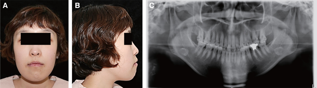
Fig. 1 Preoperative extraoral photographs. (A) Frontal view, (B) Lateral view, (C) Panoramic view.

Fig. 2 Severe erosion on entire dentition and unesthetic smile are showed on the photographs. (A) Occlusal view of maxilla, (B) Lateral view (right side), (C) Frontal view, (D) Lateral view (left side), (E) Unaesthetic smile, (F) Occlusal view of mandible.
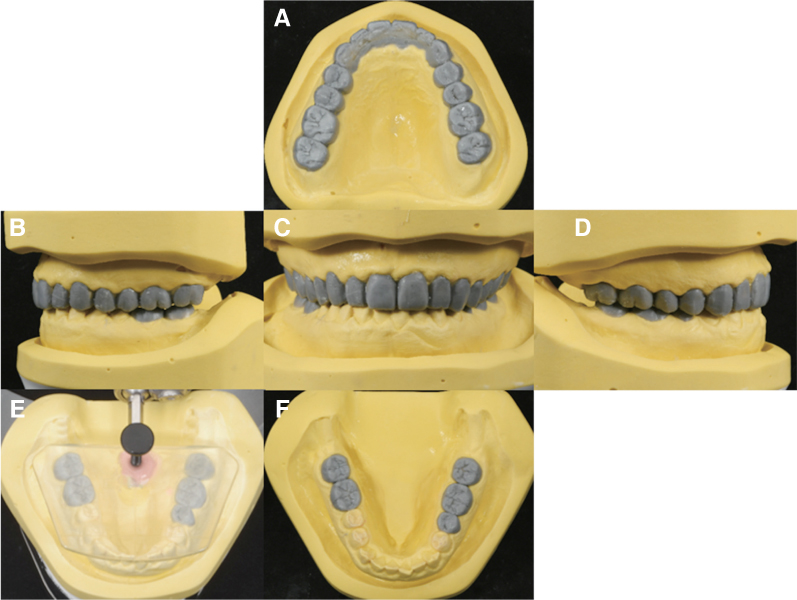
Fig. 3 Diagnostic wax-up. (A) Occlusal view of maxilla, (B) Lateral view (right side), (C) Frontal view, (D) Lateral view (left side), (E) Diagnostic wax-up using template system, (F) Occlusal view of mandible.

Fig. 4 Crown lengthening procedure (CLP) guide. (A) Facial scanning of smile, (B) Design of CLP guide, (C) CLP guide, (D) Superimposition of facial scan and CLP model, (E) Occlusal view of CLP guide, (F) Frontal view of CLP guide.

Fig. 5 Temporary prosthesis. (A) Occlusal view of maxilla, (B) Lateral view (right side), (C) Frontal view, (D) Lateral view (left side), (E) Lateral view (right side, lateral movement), (F) Occlusal view of mandible, (G) Lateral view (left side, lateral movement).
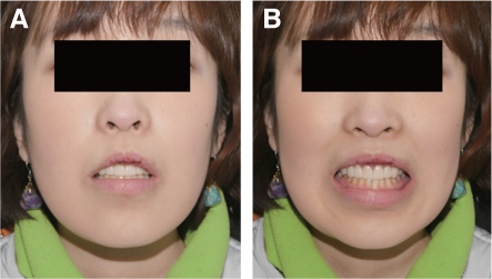
Fig. 6 (A) Frontal view of facial photograph (rest), (B) Frontal view of facial photograph (smile).
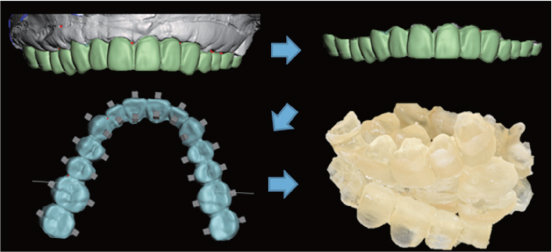
Fig. 7 Design of individual tooth tray used temporary restorations.
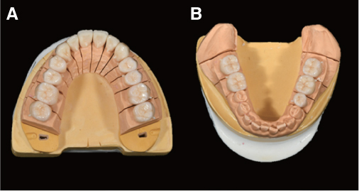
Fig. 8 (A) Definitive restorations of maxilla, (B) Definitive restorations of mandible.

Fig. 9 Definitive restoration. (A) Occlusal view of maxilla, (B) Lateral view (right side), (C) Frontal view, (D) Lateral view (left side), (E) Lateral view (right side, lateral movement), (F) Occlusal view of mandible, (G) Lateral view (left side, lateral movement).

Fig. 10 After treatment. (A) Frontal view of facial photograph (rest), (B) Frontal view of facial photograph (smile), (C) Panoramic view of definitive restorations.
Reference
-
1. Carvalho TS, Colon P, Ganss C, Huysmans MC, Lussi A, Schlueter N, Schmalz G, Shellis RP, Tveit AB, Wiegand A. Consensus report of the European Federation of Conservative Dentistry: erosive tooth wear-diagnosis and management. Clin Oral Investig. 2015; 19:1557–1561.
Article2. Bartlett DW. The role of erosion in tooth wear: aetiology, prevention and management. Int Dent J. 2005; 55:277–284.
Article3. Turner KA, Missirlian DM. Restoration of the extremely worn dentition. J Prosthet Dent. 1984; 52:467–474.
Article4. Willis FM. Features of the face involved in full denture prosthesis. Dent Cosmos. 1935; 77:851–854.5. Lan TH, Liu PH, Chou MM, Lee HE. Fracture resistance of monolithic zirconia crowns with different occlusal thicknesses in implant prostheses. J Prosthet Dent. 2016; 115:76–83.
Article6. Goodacre CJ, Campagni WV, Aquilino SA. Tooth preparations for complete crowns: an art form based on scientific principles. J Prosthet Dent. 2001; 85:363–376.
Article7. Dong JK, Jin TH, Cho HW, Oh SC. The esthetics of the smile: a review of some recent studies. Int J Prosthodont. 1999; 12:9–19.8. Rossi R, Benedetti R, Santos-Morales RI. Treatment of altered passive eruption: periodontal plastic surgery of the dentogingival junction. Eur J Esthet Dent. 2008; 3:212–223.
- Full Text Links
-
- Actions
-
Cited
- CITED
-
- Close
- Share
-
- Similar articles
-
- Full-mouth rehabilitation in a patient with severe erosion and wear using various digital tools: a case report
- Full mouth rehabilitation of partially and fully edentulous patient with crown lengthening procedure: a case report
- A case of full mouth rehabilitation with orthodontic treatment in patient with extensive tooth erosion and wear using monolithic zirconia prostheses
- Full mouth rehabilitation of the patient with severe tooth erosion using collarless porcelain fused to gold restorations: a case report
- Full mouth rehabilitation of a severely worn dentition using intraoral scanner and the CAD/CAM double scanning technique
