A multiply split femoral nerve and psoas quartus muscle
- Affiliations
-
- 1Seattle Science Foundation, Seattle, WA, USA. joei@seattlesciencefoundation.org
- 2Dental and Oral Medical Center, Kurume University School of Medicine, Kurume, Japan.
- 3Division of Gross and Clinical Anatomy, Department of Anatomy, Kurume University School of Medicine, Kurume, Japan.
- 4Department of Anatomical Sciences, St. George's University, St. George's, Grenada, West Indies.
- KMID: 2451226
- DOI: http://doi.org/10.5115/acb.2019.52.2.208
Abstract
- The femoral nerve is the largest branch of the lumbar plexus. It is normally composed of the ventral rami of spinal nerves L2 to L4. The psoas major has proximal attachments onto the T12 to L5 vertebrae and related intervertebral discs, fuses with the iliacus deep to the inguinal ligament and then attaches onto the lesser trochanter of the femur. Normally, the anatomical relationship is that the femoral nerve is located between the iliacus and psoas major. Herein, we report a case of the psoas quartus muscle related to several splits of the femoral nerve within the pelvis. Although the embryology for this is unclear, surgeons and physicians should be aware of such anatomical variants in order to better understand pain and entrapment syndromes and during surgical maneuvers in this region such as lateral transpsoas approaches to the lumbar spine.
MeSH Terms
Figure
-
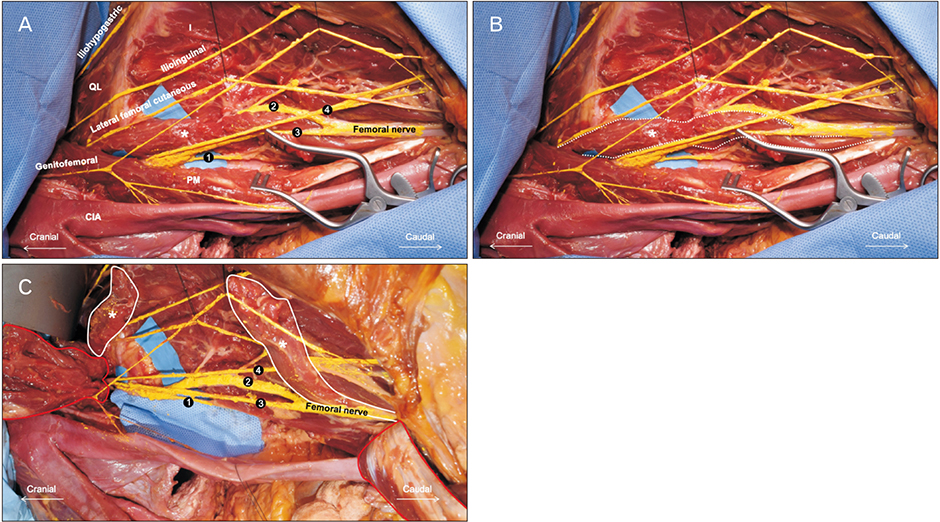
Fig. 1 (A) Photograph of the left anterior view of the case presented herein. The psoas quartus (asterisk) and split femoral nerve is shown. (B) The psoas quartus is outlined by the white dotted line. (C) After cutting and reflecting the psoas quartus (asterisks and white solid lines) and the psoas major (red solid lines) the following structures are shown. CIA, common iliac artery; I, iliacus; PM, psoas major; QL, quadratus lumborum; 1, the most medial branch of the FN; 2, the second branch of the FN; 3, the third branch of the FN; 4, the most lateral branch.
Cited by 1 articles
-
The ascending branch of the lateral circumflex femoral artery penetrating the posterior division of the femoral nerve
Hankyu Kim, Yong Seok Nam, Yi-Suk Kim
Anat Cell Biol. 2021;54(1):124-127. doi: 10.5115/acb.20.237.
Reference
-
1. Standring S. Gray's anatomy: the anatomical basis of clinical practice. 41st ed. New York: Elsevier;2016. p. 1094–1095.2. Clarkson RD, Rainy H. Unusual arrangement of the psoas muscle. J Anat Physiol. 1889; 23(Pt 3):504–506.3. Jelev L, Shivarov V, Surchev L. Bilateral variations of the psoas major and the iliacus muscles and presence of an undescribed variant muscle: accessory iliopsoas muscle. Ann Anat. 2005; 187:281–286.4. Khalid S, Iwanaga J, Loukas M, Tubbs RS. Split femoral nerve due to psoas tertius muscle: a review with other cases of variant muscles traversing the femoral nerve. Cureus. 2017; 9:e1555.
Article5. Tubbs RS, Oakes WJ, Salter EG. The psoas quartus muscle. Clin Anat. 2006; 19:678–680.
Article6. Vazquez MT, Murillo J, Maranillo E, Parkin IG, Sanudo J. Femoral nerve entrapment: a new insight. Clin Anat. 2007; 20:175–179.7. Kirchmair L, Lirk P, Colvin J, Mitterschiffthaler G, Moriggl B. Lumbar plexus and psoas major muscle: not always as expected. Reg Anesth Pain Med. 2008; 33:109–114.
Article8. Anloague PA, Huijbregts P. Anatomical variations of the lumbar plexus: a descriptive anatomy study with proposed clinical implications. J Man Manip Ther. 2009; 17:e107–e114.
Article9. Perry CP. Peripheral neuropathies and pelvic pain: diagnosis and management. Clin Obstet Gynecol. 2003; 46:789–796.
Article10. Tubbs RS, Salter EG, Wellons JC 3rd, Blount JP, Oakes WJ. Anatomical landmarks for the lumbar plexus on the posterior abdominal wall. J Neurosurg Spine. 2005; 2:335–338.
Article11. Mahan MA, Sanders LE, Guan J, Dailey AT, Taylor W, Morton DA. Anatomy of psoas muscle innervation: cadaveric study. Clin Anat. 2017; 30:479–486.
Article12. Aichroth P, Rowe-Jones DC. Iliacus compartment compression syndrome. Br J Surg. 1971; 58:833–834.
Article13. Spratt JD, Logan BM, Abrahams PH. Variant slips of psoas and iliacus muscles, with splitting of the femoral nerve. Clin Anat. 1996; 9:401–404.
Article14. Moore KL, Persaud TV, Torchia MG. The developing human. 10th ed. Philadelphia: Elsevier;2016. p. 357–358. p. 36715. Sakamoto J, Manabe Y, Oyamada J, Kataoka H, Nakano J, Saiki K, Okamoto K, Tsurumoto T, Okita M. Anatomical study of the articular branches innervated the hip and knee joint with reference to mechanism of referral pain in hip joint disease patients. Clin Anat. 2018; 31:705–709.
Article
