Outcomes of Anti-vascular Endothelial Growth Factor Treatment for Foveal Serous Retinal Detachment Associated with Inferior Staphyloma
- Affiliations
-
- 1Department of Ophthalmology, Jeju National University Hospital, Jeju National University School of Medicine, Jeju, Korea.
- 2Department of Ophthalmology, Seoul National University Hospital, Seoul National University College of Medicine, Seoul, Korea. hgonyu@snu.ac.kr
- KMID: 2448862
- DOI: http://doi.org/10.3341/kjo.2018.0125
Abstract
- PURPOSE
To evaluate the efficacy of anti-vascular endothelial growth factor (VEGF) treatment of eyes with foveal serous retinal detachment (SRD) associated with inferior staphyloma and to investigate choroidal thickness changes following anti-VEGF therapy.
METHODS
In this observational case series, eyes with inferior staphyloma accompanied by foveal SRD were treated with a single intravitreal anti-VEGF injection, followed by further injections as needed. Changes in height and width of subretinal fluid (SRF) and visual acuity after treatment were assessed. Choroidal thickness was measured at the subfovea, 1.5 mm superior and inferior to the fovea using enhanced depth imaging optical coherence tomography at baseline and 1 month after initial anti-VEGF therapy.
RESULTS
Six eyes from six patients were included. One month after the initial injection, the mean SRF height and width had decreased significantly from 112.5 ± 40.1 to 44.5 ± 48.7 µm (p = 0.046) and from 1,401.8 ± 627.3 to 690.7 ± 634.7 µm (p = 0.028), respectively. Mean choroidal thickness at the superior point decreased from 218.7 ± 59.3 to 200.5 ± 61.0 µm (p = 0.046). SRF resolved completely in three of the six eyes (50%) with a mean of 6.8 ± 5.9 injections (range, 1 to 15). All eyes experienced at least one recurrence of exudation, at a mean interval of 4.8 months. Mean visual acuity improvement was 0.17 logarithm of the minimum angle of resolution units at a mean of 28.7 months follow-up.
CONCLUSIONS
Anti-VEGF therapy resulted in an SRF decrease and modest visual improvement in eyes with foveal SRD associated with inferior staphyloma. Reduction in superior choroidal thickness appeared to contribute to the clinical improvements that were observed.
Keyword
MeSH Terms
Figure
-
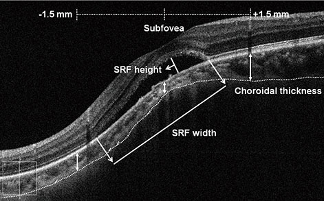
Fig. 1 An enhanced depth imaging optical coherence tomography image demonstrating the morphologic parameters measured in this study. The dotted line indicates the posterior edge of the choroid, and the white double arrows indicate the choroidal thickness. SRF = subretinal fluid.
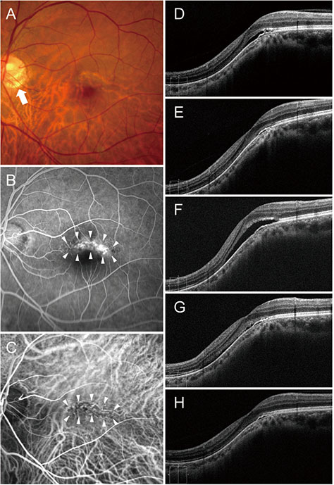
Fig. 2 A 52-year-old man (case 1) with foveal serous retinal detachment associated with inferior staphyloma. (A) Fundus photograph revealed inferior staphyloma and inferior peripapillary crescent (arrow). (B) Fluorescein angiography showed a belt-shaped area of granular hyperfluorescence (arrowheads) corresponding to the border of the inferior staphyloma. (C) Indocyanine green angiography demonstrated a hypofluorescent band (arrowheads). (D) Baseline spectral-domain optical coherence tomography showed foveal serous retinal detachment. (E) After two bevacizumab injections, subretinal fluid (SRF) resolved completely. (F) The patient experienced a mild recurrence and resolution after the injection, and he received four bevacizumab injections over a period of 2 years. Thereafter, SRF recurred. (G) After two more injections, SRF decreased. (H) Seven additional bevacizumab injections were performed with repeated recurrence and resolution, and no SRF was observed at the last visit.
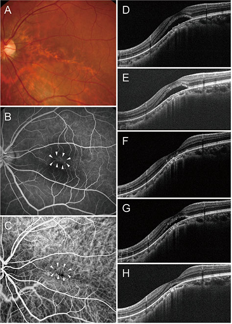
Fig. 3 A 41-year-old man (case 2) with foveal serous retinal detachment (SRD) associated with inferior staphyloma. (A) Fundus photograph showed inferior staphyloma with a superior border across the macula. (B) Fluorescein angiography revealed multiple pinpoint staining (arrowheads). (C) Indocyanine green angiography demonstrated subtle hypofluorescence (arrowheads). (D) Baseline spectral-domain optical coherence tomography showed foveal SRD. (E) After three bevacizumab injections, subretinal fluid (SRF) decreased slightly, but persisted. (F) Complete resolution of SRF was noted after three more bevacizumab injections. (G) SRD recurred 3 months later. (H) After one more injection, SRF decreased, but remained present.
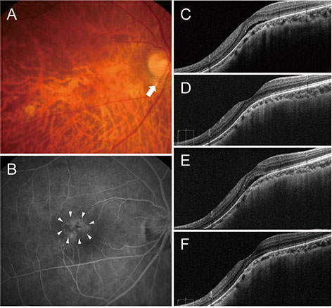
Fig. 4 A 68-year-old woman (case 3) with foveal serous retinal detachment associated with inferior staphyloma. (A) Fundus photograph showed inferior staphyloma and inferior peripapillary crescent (arrow). (B) Fluorescein angiography revealed multiple pinpoint staining (arrowheads). (C) Baseline spectral-domain optical coherence tomography demonstrated foveal serous retinal detachment. (D) After one ranibizumab injection, the subretinal fluid (SRF) resolved completely. (E) The patient experienced repeated mild recurrence and resolution after the injection, and she received nine ranibizumab injections over a period of 3 years. Thereafter, a small amount of SRF accumulated again. (F) After two additional injections for 8 months, no SRF was observed at the last visit.
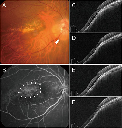
Fig. 5 A 59-year-old woman (case 4) with foveal serous retinal detachment associated with inferior staphyloma. (A) Fundus photograph showed inferior staphyloma and inferior peripapillary crescent (arrow). (B) Fluorescein angiography revealed multiple pinpoint staining (arrowheads). (C) Baseline spectral-domain optical coherence tomography demonstrated foveal serous retinal detachment. (D) After one bevacizumab injection, subretinal fluid (SRF) resolved completely. (E) SRF increased 10 months later. (F) After one more bevacizumab injection, the SRF was completely absorbed.
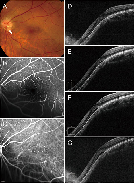
Fig. 6 A 36-year-old man (case 5) with foveal serous retinal detachment (SRD) associated with inferior staphyloma. (A) Fundus photograph showed inferior staphyloma and inferior peripapillary crescent (arrow). (B) There was no evidence of leakage or staining on fluorescein angiography. (C) Indocyanine green angiography demonstrated subtle hypofluorescence (arrowheads). (D) Baseline spectral-domain optical coherence tomography showed foveal SRD. (E) One aflibercept injection decreased the amount of subretinal fluid (SRF) slightly, but the SRF persisted. (F) The SRF had increased 4 months later. (G) The patient declined further injections. At the final visit, the foveal SRD remained.
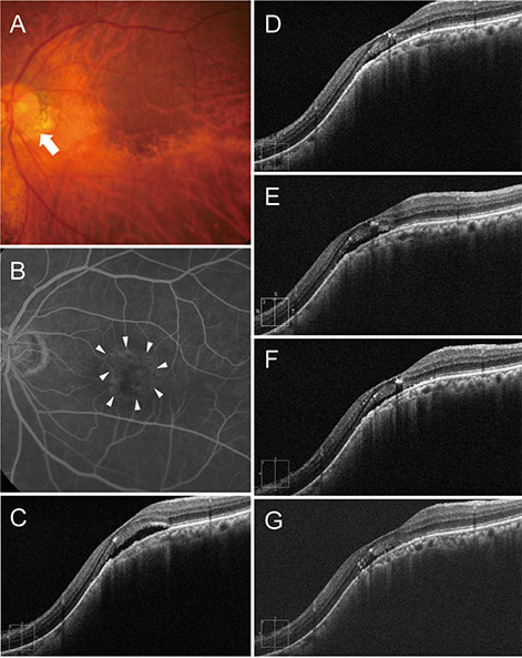
Fig. 7 A 76-year-old woman (case 6) with foveal serous retinal detachment associated with inferior staphyloma. (A) Fundus photograph showed inferior staphyloma and inferior peripapillary crescent (arrow). (B) Fluorescein angiography revealed multiple pinpoint staining (arrowheads). (C) Baseline spectral-domain optical coherence tomography showed foveal serous retinal detachment. (D) Two bevacizumab injections reduced the amount of subretinal fluid (SRF), but SRF was still present. (E) SRF had increased 2 months later. (F) After one more bevacizumab injection, SRF decreased slightly, but remained. (G) The patient declined further injections. At the final visit, little SRF remained.
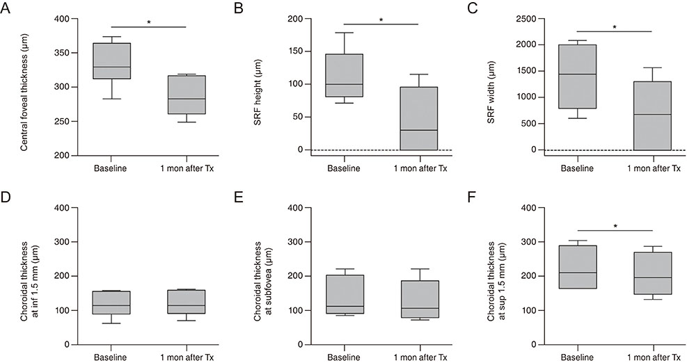
Fig. 8 Comparison of measurements at baseline and 1 month after the initial anti-vascular endothelial growth factor injection. (A) Central foveal thickness, (B) height and (C) width of subretinal fluid (SRF), and (D) choroidal thicknesses measured 1.5 mm inferior to the fovea, (E) at the subfovea, and (F) 1.5 mm superior to the fovea. Box plots indicate median values with 5th and 95th percentiles. Tx = treatment. *p < 0.05 by Wilcoxon signed rank test.
Reference
-
1. Curtin BJ. The posterior staphyloma of pathologic myopia. Trans Am Ophthalmol Soc. 1977; 75:67–86.2. Apple DJ, Rabb MF, Walsh PM. Congenital anomalies of the optic disc. Surv Ophthalmol. 1982; 27:3–41.
Article3. Tosti G. Serous macular detachment and tilted disc syndrome. Ophthalmology. 1999; 106:1453–1455.
Article4. Leys AM, Cohen SY. Subretinal leakage in myopic eyes with a posterior staphyloma or tilted disk syndrome. Retina. 2002; 22:659–665.
Article5. Theodossiadis PG, Grigoropoulos V, Emfietzoglou J, Theodossiadis GP. Optical coherence tomography study of tilted optic disk associated with macular detachment. Graefes Arch Clin Exp Ophthalmol. 2006; 244:122–124.
Article6. Nakanishi H, Tsujikawa A, Gotoh N, et al. Macular complications on the border of an inferior staphyloma associated with tilted disc syndrome. Retina. 2008; 28:1493–1501.
Article7. Cohen SY, Quentel G, Guiberteau B, et al. Macular serous retinal detachment caused by subretinal leakage in tilted disc syndrome. Ophthalmology. 1998; 105:1831–1834.
Article8. Brown DM, Kaiser PK, Michels M, et al. Ranibizumab versus verteporfin for neovascular age-related macular degeneration. N Engl J Med. 2006; 355:1432–1444.
Article9. Rosenfeld PJ, Brown DM, Heier JS, et al. Ranibizumab for neovascular age-related macular degeneration. N Engl J Med. 2006; 355:1419–1431.
Article10. Ikuno Y, Sayanagi K, Soga K, et al. Intravitreal bevacizumab for choroidal neovascularization attributable to pathological myopia: one-year results. Am J Ophthalmol. 2009; 147:94–100.
Article11. Silva RM, Ruiz-Moreno JM, Rosa P, et al. Intravitreal ranibizumab for myopic choroidal neovascularization: 12-month results. Retina. 2010; 30:407–412.12. Milani P, Pece A, Pierro L, et al. Bevacizumab for macular serous neuroretinal detachment in tilted disk syndrome. J Ophthalmol. 2010; 2010:970580.
Article13. Donati MC, Miele A, Abbruzzese G, et al. Treatment of macular serous neuroretinal detachment in tilted disk syndrome: report of 3 cases. Eur J Ophthalmol. 2013; 23:267–270.
Article14. Spaide RF, Koizumi H, Pozzoni MC. Enhanced depth imaging spectral-domain optical coherence tomography. Am J Ophthalmol. 2008; 146:496–500.
Article15. Hirata M, Tsujikawa A, Matsumoto A, et al. Macular choroidal thickness and volume in normal subjects measured by swept-source optical coherence tomography. Invest Ophthalmol Vis Sci. 2011; 52:4971–4978.
Article16. Yamagishi T, Koizumi H, Yamazaki T, Kinoshita S. Choroidal thickness in inferior staphyloma associated with posterior serous retinal detachment. Retina. 2012; 32:1237–1242.
Article17. Ellabban AA, Tsujikawa A, Matsumoto A, et al. Macular choroidal thickness measured by swept source optical coherence tomography in eyes with inferior posterior staphyloma. Invest Ophthalmol Vis Sci. 2012; 53:7735–7745.
Article18. Maruko I, Iida T, Sugano Y, et al. Morphologic choroidal and scleral changes at the macula in tilted disc syndrome with staphyloma using optical coherence tomography. Invest Ophthalmol Vis Sci. 2011; 52:8763–8768.
Article19. Murakami T, Felinski EA, Antonetti DA. Occludin phosphorylation and ubiquitination regulate tight junction trafficking and vascular endothelial growth factor-induced permeability. J Biol Chem. 2009; 284:21036–21046.
Article20. Nicoletti VG, Nicoletti R, Ferrara N, et al. Diabetic patients and retinal proliferation: an evaluation of the role of vascular endothelial growth factor (VEGF). Exp Clin Endocrinol Diabetes. 2003; 111:209–214.
Article21. Saenz-de-Viteri M, Fernandez-Robredo P, Hernandez M, et al. Single- and repeated-dose toxicity study of bevacizumab, ranibizumab, and aflibercept in ARPE-19 cells under normal and oxidative stress conditions. Biochem Pharmacol. 2016; 103:129–139.
Article22. Hirano Y, Yasukawa T, Tsukada A, et al. Resolution of exudative changes refractory to ranibizumab after aflibercept injections at the margin of inferior staphyloma in tilted disc syndrome. Ophthalmic Surg Lasers Imaging Retina. 2015; 46:384–386.
Article
- Full Text Links
-
- Actions
-
Cited
- CITED
-
- Close
- Share
-
- Similar articles
-
- A Case of Retinal Detachment with Equatorial Scleral Staphyloma
- A Case of Retinal Detachment with Equatorial Scleral Staphyloma
- A Case of Polypoidal Choroidal Vasculopathy and Serous Retinal Detachment in a Bilateral Dome-shaped Macula
- Review and update for central serous chorioretinopathy
- Anti-Vascular Endothelial Growth Factor Therapy for Choroidal Neovascularization Secondary to Optic Nerve Head Drusen
