J Korean Ophthalmol Soc.
2019 Dec;60(12):1369-1373. 10.3341/jkos.2019.60.12.1369.
Anti-Vascular Endothelial Growth Factor Therapy for Choroidal Neovascularization Secondary to Optic Nerve Head Drusen
- Affiliations
-
- 1Nune Eye Hospital, Seoul, Korea. ophkh@hanmail.net
- KMID: 2466198
- DOI: http://doi.org/10.3341/jkos.2019.60.12.1369
Abstract
- PURPOSE
To describe a patient with optic nerve head drusen who showed improved retinal hemorrhage and visual acuity following intravitreal anti-vascular endothelial growth factor (bevacizumab) injection.
CASE SUMMARY
A 53-year-old woman with no underlying disease presented with sudden vision loss in her left eye. Her best-corrected visual acuity was 1.0 in the right eye and 0.8 in the left eye; the intraocular pressure was 15 mmHg in both eyes. Anterior segment examination revealed no abnormal findings. Fundus examination showed subretinal hemorrhage and serous retinal detachment in the left eye. Fluorescein angiography and indocyanine green fundus examination revealed hyperfluorescence near the optic disc. The patient was diagnosed with choroidal neovascularization of the left eye secondary to optic nerve head drusen; intravitreal bevacizumab injection was then performed. Three weeks later, the patient showed improved retinal hemorrhage.
CONCLUSIONS
Intravitreal anti-vascular endothelial growth factor injection may be an effective treatment for choroidal neovascularization associated with optic nerve head drusen.
Keyword
MeSH Terms
-
Bevacizumab
Choroid*
Choroidal Neovascularization*
Endothelial Growth Factors*
Female
Fluorescein Angiography
Hemorrhage
Humans
Indocyanine Green
Intraocular Pressure
Intravitreal Injections
Middle Aged
Optic Disk*
Optic Nerve*
Retinal Detachment
Retinal Hemorrhage
Visual Acuity
Bevacizumab
Endothelial Growth Factors
Indocyanine Green
Figure
-
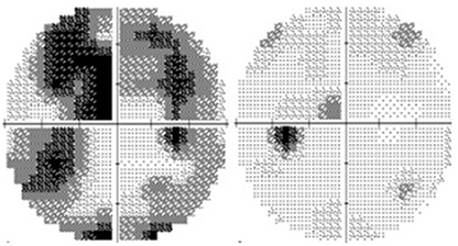
Figure 1 Visual field tests of both eyes at the first visit.
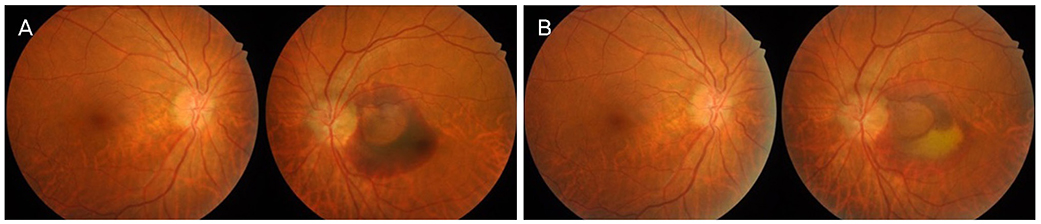
Figure 2 Fundus photography of both eyes. (A) Pre-treatment and (B) post-treatment fundus photography at 3 weeks after intravitrealbevacizumab injection.
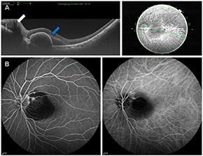
Figure 3 Optical coherence tomography(OCT) and retinal angiograms of the lefteye. (A) A hyperreflective subretinal masslikelesion (white arrow) is buried at opticdisc and subretinal fluid extended fromthe optic nerve head to the macula andpigment epithelial detachment (blue arrow)is observed on OCT. (B) Fluoresceinand Indocyanine green angiogram revealedround small hyperfluorescencewithin the hypofluorescence area of subretinalhemorrhage.
Reference
-
1. Auw-Haedrich C, Staubach F, Witschel H. Optic disk drusen. Surv Ophthalmol. 2002; 47:515–532.2. Palmer E, Gale J, Crowston JG, Wells AP. Optic nerve head drusen: an update. Neuroophthalmology. 2018; 42:367–384.3. Romero J, Sowka J, Shechtman D. Hemorrhagic complications of optic disc drusen and available treatment options. Optometry. 2008; 79:496–500.4. Kurz-Levin MM, Landau K. A comparison of imaging techniques for diagnosing drusen of the optic nerve head. Arch Ophthalmol. 1999; 117:1045–1049.5. Lee KM, Woo SJ, Hwang JM. Differentiation of optic nerve head drusen and optic disc edema with spectral-domain optical coherence tomography. Ophthalmology. 2011; 118:971–977.6. Seo DR, Park SH. Optical coherence tomography findings of optic nerve head drusen in children and adolescents. J Korean Ophthalmol Soc. 2015; 56:1446–1453.7. Grippo TM, Shihadeh WA, Schargus M, et al. Optic nerve head drusen and visual field loss in normotensive and hypertensive eyes. J Glaucoma. 2008; 17:100–104.8. Choi WG, Jang JH. A case of optic disc hemorrhage associated with buried optic disc nerve head drusen. J Korean Ophthalmol Soc. 2014; 55:1099–1105.9. Duncan JE, Freedman SF, El-Dairi MA. The incidence of neovascular membranes and visual field defects from optic nerve head drusen in children. J AAPOS. 2016; 20:44–48.10. Delyfer MN, Rougier MB, Fourmaux E, et al. Laser photocoagulation for choroidal neovascular membrane associated with optic disc drusen. Acta Ophthalmol Scand. 2004; 82:236–238.11. Silva R, Torrent T, Loureiro R, et al. Bilateral CNV associated with optic nerve drusen treated with photodynamic therapy with verteporfin. Eur J Ophthalmol. 2004; 14:434–437.12. Mateo C, Moreno JG, Lechuga M, et al. Surgical removal of peripapillary choroidal neovascularization associated with optic nerve drusen. Retina. 2004; 24:739–745.13. Knape RM, Zavaleta EM, Clark CL 3rd, et al. Intravitreal bevacizumab treatment of bilateral peripapillary choroidal neovascularization from optic nerve head drusen. J AAPOS. 2011; 15:87–90.14. Delas B, Almudí L, Carreras A, Asaad M. Bilateral choroidal neovascularization associated with optic nerve head drusen treated by antivascular endothelial growth factor therapy. Clin Ophthalmol. 2012; 6:225–230.15. Saffra NA, Reinherz BJ. Peripapillary choroidal neovascularization associated with optic nerve head drusen treated with anti-VEGF agents. Case Rep Ophthalmol. 2015; 6:51–55.
- Full Text Links
-
- Actions
-
Cited
- CITED
-
- Close
- Share
-
- Similar articles
-
- Treatment of Exudative Age-Related Macular Degeneration
- The Role of VEGF and Bruch's Membrane in the Development Choroidal Neovascularization
- Bilateral Optic Disc Drusen Mimicking Papilledema
- Optic Nerve Head Drusen Mimicking Optic Nerve Tumor
- The Effect of TGF-beta 2 and TNF-alpha on the Migration of Choroidal Endothelial Cells Doheny Eye Institute
