Review and update for central serous chorioretinopathy
- Affiliations
-
- 1Department of ophthalmology, Hanyang University Guri Hospital, Gyeonggi-do, Korea. syu2000@hanmail.net
- KMID: 2384002
- DOI: http://doi.org/10.7599/hmr.2017.37.1.10
Abstract
- Central serous chorioretinopathy (CSC) is an eye disease that causes serous retinal detachment in the posterior pole of the retina. The pathogenesis of CSC is not fully understood and various systemic factors have been reported to be associated with CSC. Recently, with the advent of advanced imaging techniques, novel imaging findings for CSC have been reported and the understanding of CSC has increased further. Moreover, in addition to conventional treatment for CSC, new treatment modalities such as photodynamic therapy, anti-vascular endothelial growth factor or subthreshold laser therapy, have emerged. In this article, an overall review and update of CSC, particularly focusing on new imaging findings and treatments, will be discussed.
Keyword
MeSH Terms
Figure
-
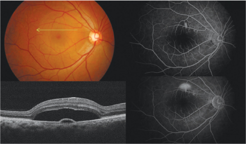
Fig. 1 Representative case of acute central serous chorioretinopathy. Top left, fundus photography shows discrete, clear and serous elevation in central area. Green arrow indicates the area of section on optical coherence tomography. Bottom left, optical coherence tomography shows subretinal fluid, thickened posterior surface of detached retina and semicircular pigment epithelial detachment. Right, fluorescein angiography shows smoke-stack like dye leakage (Top; early phase, bottom; late phase).
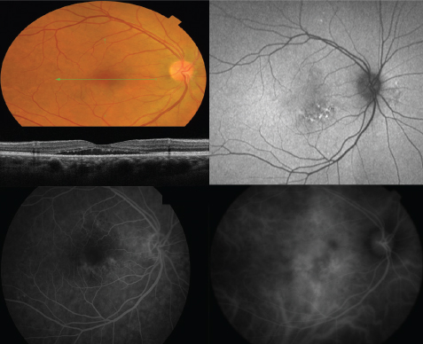
Fig. 2 Representative case of chronic central serous chorioretinopathy. Upper left, fundus photography shows subtle serous elevation in central area. Green arrow indicates the area of section on optical coherence tomography. Middle left, optical coherence shows shallow subretinal fluid, thinned posterior surface of detached retina, subretinal hype-reflective foci. Top right, fundus autofluorscence shows heterogeneous patterns of hyper-fluorescence. Bottom left, fluorescein angiography (mid-phase) shows multifocal leakages as patches of granular hyperfluorescence. Bottom right, indocyanine green angiography (late phase) shows hyperfluorescent areas with dilating choroidal vessels (choroidal vascular hyperpermeability).
Cited by 1 articles
-
Current update in diverse diseases
Seong-Ho Koh
Hanyang Med Rev. 2017;37(1):1-1. doi: 10.7599/hmr.2017.37.1.1.
Reference
-
1. Spaide RF, Campeas L, Haas A, Yannuzzi LA, Fisher YL, Guyer DR, et al. Central serous chorioretinopathy in younger and older adults. Ophthalmology. 1996; 103:2070–2079.
Article2. Von Graefe A. Ueber centrale recidivierende retinitis. Graefes Arch Clin Exp Ophthalmol. 1866; 12:211–215.3. Maumenee A. Clinical manifestations (Symposium: macular diseases). Trans Am Acad Ophthalmol Otolaryngol. 1965; 69:605–613.4. Donald J, Gass M. Pathogenesis of disciform detachment of the neuroepithelium: II. Idiopathic central serous choroidopathy. Am J Ophthalmol. 1967; 63:587/15–615/43.5. Kitzmann AS, Pulido JS, Diehl NN, Hodge DO, Burke JP. The incidence of central serous chorioretinopathy in Olmsted County, Minnesota, 1980–2002. Ophthalmology. 2008; 115:169–173.
Article6. Haimovici R, Koh S, Gagnon DR, Lehrfeld T, Wellik S. Group CSCCCS. Risk factors for central serous chorioretinopathy: a case–control study. Ophthalmology. 2004; 111:244–249.7. Chan WM, Lai TY, Tano Y, Liu DT, Li KK, Lam DS. Photodynamic therapy in macular diseases of Asian populations: when East meets West. Jpn J Ophthalmol. 2006; 50:161–169.
Article8. Balo K, Mihluedo H. [Idiopathic central serous chorioretinopathy: two case reports observed in Togo]. Med Trop (Mars). 1996; 56:381–383.9. Desai UR, Alhalel AA, Campen TJ, Schiffman RM, Edwards PA, Jacobsen GR. Central serous chorioretinopathy in African Americans. J Natl Med Assoc. 2003; 95:553–559.10. Liu B, Deng T, Zhang J. RISK FACTORS FOR CENTRAL SEROUS CHORIORETINOPATHY: A Systematic Review and Meta-Analysis. Retina. 2016; 36:9–19.11. Carvalho-Recchia CA, Yannuzzi LA, Negrao S, Spaide RF, Freund KB, Rodriguez-Coleman H, et al. Corticosteroids and central serous chorioretinopathy. Ophthalmology. 2002; 109:1834–1837.
Article12. Khairallah M, Kahloun R, Tugal-Tutkun I. Central serous chorioretinopathy, corticosteroids, and uveitis. Ocul Immunol Inflamm. 2012; 20:76–85.
Article13. Chung H, Kim KH, Kim JG, Lee SY, Yoon YH. Retinal complications in patients with solid organ or bone marrow transplantations. Transplantation. 2007; 83:694–699.
Article14. Mayo GL, Tolentino MJ. Images in clinical medicine. Central serous chorioretinopathy in pregnancy. N Engl J Med. 2005; 353:e6.15. Yannuzzi LA. Type A behavior and central serous chorioretinopathy. Trans Am Ophthalmol Soc. 1986; 84:799.
Article16. Tittl MK, Spaide RF, Wong D, Pilotto E, Yannuzzi LA, Fisher YL, et al. Systemic findings associated with central serous chorioretinopathy. Am J Ophthalmol. 1999; 128:63–68.
Article17. Mansuetta CC, Mason JO, Swanner J, Feist RM, White MF, Thomley ML, et al. An association between central serous chorioretinopathy and gastroesophageal reflux disease. Am J Ophthalmol. 2004; 137:1096–1100.
Article18. Cotticelli L, Borrelli M, d'Alessio A, Menzione M, Villani A, Piccolo G, et al. Central serous chorioretinopathy and Helicobacter pylori. Eur J Ophthalmol. 2006; 16:274–278.
Article19. Daruich A, Matet A, Dirani A, Bousquet E, Zhao M, Farman N, et al. Central serous chorioretinopathy: Recent findings and new physiopathology hypothesis. Prog Retin Eye Res. 2015; 48:82–118.
Article20. Ross A, Ross AH, Mohamed Q. Review and update of central serous chorioretinopathy. Curr Opin Ophthalmol. 2011; 22:166–173.
Article21. Framme C, Walter A, Gabler B, Roider J, Sachs HG, Gabel VP. Fundus autofluorescence in acute and chronic-recurrent central serous chorioretinopathy. Acta Ophthalmol Scand. 2005; 83:161–167.
Article22. Imamura Y, Fujiwara T, Margolis R, Spaide RF. Enhanced depth imaging optical coherence tomography of the choroid in central serous chorioretinopathy. Retina. 2009; 29:1469–1473.
Article23. Montero J, Ruiz-Moreno J. Optical coherence tomography characterisation of idiopathic central serous chorioretinopathy. Br J Ophthalmol. 2005; 89:562–564.
Article24. Yoshioka H, Katsume Y, Akune H. Experimental Central Serous Chorioretinopathy in Monkey Eyes: Fluorescein Angiographic Findings. Ophthalmologica. 1982; 185:168–178.
Article25. Sakaue M, Hoffman BB. Glucocorticoids induce transcription and expression of the alpha 1B adrenergic receptor gene in DTT1 MF-2 smooth muscle cells. J Clin Invest. 1991; 88:385.
Article26. Caccavale A, Romanazzi F, Imparato M, Negri A, Morano A, Ferentini F. Central serous chorioretinopathy: a pathogenetic model. Clin Ophthalmol. 2011; 5:239.
Article27. Verma L, Purohit A, Tewari H, Biswas N, Talwar D, Jhingan S, et al. A study of endogenous cortisol profile in patients with central serous retinopathy with single and multiple leaks. Indian J Pharmacol. 2001; 33:96–99.28. Gilbert CM, Owens SL, Smith PD, Fine SL. Long-term follow-up of central serous chorioretinopathy. Br J Ophthalmol. 1984; 68:815–820.
Article29. Yannuzzi LA, Shakin JL, Fisher YL, Altomonte MA. Peripheral retinal detachments and retinal pigment epithelial atrophic tracts secondary to central serous pigment epitheliopathy. Ophthalmology. 1984; 91:1554–1572.
Article30. Jalkh AE, Jabbour N, Avila MP, Trempe CL, Schepens CL. Retinal pigment epithelium decompensation. I. Clinical features and natural course. Ophthalmology. 1984; 91:1544–1548.31. Iida T, Yannuzzi LA, Spaide RF, Borodoker N, Carvalho CA, Negrao S. Cystoid macular degeneration in chronic central serous chorioretinopathy. Retina. 2003; 23:1–7. quiz 137-8.
Article32. Gäckle HC, Lang GE, Freißler KA, Lang GK. Clinical, fluorescein angiographic and demographic aspects in central serous chorioretinopathy. Ophthalmologe. 1998; 95:529–533.
Article33. Yamada K, Hayasaka S, Setogawa T. Fluorescein-angiographic patterns in patients with central serous chorioretinopathy at the initial visit. Ophthalmologica. 1992; 205:69–76.
Article34. Burumcek E, Mudun A, Karacorlu S, Arslan MO. Laser photocoagulation for persistent central serous retinopathy: results of long-term follow-up. Ophthalmology. 1997; 104:616–622.
Article35. Prünte C. Indocyanine green angiographic findings in central serous chorioretinopathy. Int Ophthalmol. 1995; 19:77–82.
Article36. Iida T, Kishi S, Hagimura N, Shimizu K. Persistent and bilateral choroidal vascular abnormalities in central serous chorioretinopathy. Retina. 1999; 19:508–512.
Article37. Spaide RF. Enhanced depth imaging optical coherence tomography of retinal pigment epithelial detachment in age-related macular degeneration. Am J Ophthalmol. 2009; 147:644–652.
Article38. Hirata M, Tsujikawa A, Matsumoto A, Hangai M, Ooto S, Yamashiro K, et al. Macular choroidal thickness and volume in normal subjects measured by swept-source optical coherence tomography. Invest Ophthalmol Vis Sci. 2011; 52:4971–4978.
Article39. Nicholson B, Noble J, Forooghian F, Meyerle C. Central serous chorioretinopathy: update on pathophysiology and treatment. Surv Ophthalmol. 2013; 58:103–126.
Article40. Song IS, Shin YU, Lee BR. Time-periodic characteristics in the morphology of idiopathic central serous chorioretinopathy evaluated by volume scan using spectral-domain optical coherence tomography. Am J Ophthalmol. 2012; 154:366–375.
Article41. Maruko I, Iida T, Sugano Y, Ojima A, Sekiryu T. Subfoveal choroidal thickness in fellow eyes of patients with central serous chorioretinopathy. Retina. 2011; 31:1603–1608.
Article42. Eandi CM, Ober M, Iranmanesh R, Peiretti E, Yannuzzi LA. Acute central serous chorioretinopathy and fundus autofluorescence. Retina. 2005; 25:989–993.
Article43. Lee WJ, Lee JH, Lee BR. Fundus autofluorescence imaging patterns in central serous chorioretinopathy according to chronicity. Eye (Lond). 2016; 30:1336–1342.
Article44. Yannuzzi LA. Central serous chorioretinopathy: a personal perspective. Am J Ophthalmol. 2010; 149:361–363.e1.
Article45. Sharma T, Shah N, Rao M, Gopal L, Shanmugam MP, Gopalakrishnan M, et al. Visual outcome after discontinuation of corticosteroids in atypical severe central serous chorioretinopathy. Ophthalmology. 2004; 111:1708–1714.
Article46. Leaver P, Williams C. Argon laser photocoagulation in the treatment of central serous retinopathy. Br J Ophthalmol. 1979; 63:674–677.
Article47. Robertson DM. Argon laser photocoagulation treatment in central serous chorioretinopathy. Ophthalmology. 1986; 93:972–974.
Article48. Gilbert CM, Owens SL, Smith PD, Fine SL. Long-term follow-up of central serous chorioretinopathy. Br J Ophthalmol. 1984; 68:815–820.
Article49. Salehi M, Wenick AS, Law HA, Evans JR, Gehlbach P. Interventions for central serous chorioretinopathy: a network meta-analysis. Cochrane Database Syst Rev. 2015; CD011841.
Article50. Taban M, Boyer DS, Thomas EL, Taban M. Chronic central serous chorioretinopathy: photodynamic therapy. Am J Ophthalmol. 2004; 137:1073–1080.
Article51. Lim JW, Kang SW, Kim Y-T, Chung SE, Lee SW. Comparative study of patients with central serous chorioretinopathy undergoing focal laser photocoagulation or photodynamic therapy. Br J Ophthalmol. 2011; 95:514–517. DOI: bjo.2010.182121.
Article52. Inoue R, Sawa M, Tsujikawa M, Gomi F. Association between the efficacy of photodynamic therapy and indocyanine green angiography findings for central serous chorioretinopathy. Am J Ophthalmol. 2010; 149:441–446.e2.
Article53. Shin JY, Woo SJ, Yu HG, Park KH. Comparison of efficacy and safety between half-fluence and full-fluence photodynamic therapy for chronic central serous chorioretinopathy. Retina. 2011; 31:119–126.
Article54. Reibaldi M, Cardascia N, Longo A, Furino C, Avitabile T, Faro S, et al. Standard-fluence versus low-fluence photodynamic therapy in chronic central serous chorioretinopathy: a nonrandomized clinical trial. Am J Ophthalmol. 2010; 149:307–315.e2.
Article55. Nicoló M, Eandi CM, Alovisi C, Grignolo FM, Traverso CE, Musetti D, et al. Half-fluence versus half-dose photodynamic therapy in chronic central serous chorioretinopathy. Am J Ophthalmol. 2014; 157:1033–1037.e2.
Article56. Alkin Z, Ozkaya A, Agca A, Yazici AT, Demirok A. Early visual and morphologic changes after half-fluence photodynamic therapy in chronic central serous chorioretinopathy. J Ocul Pharmacol Ther. 2014; 30:359–365.
Article57. Roisman L, Magalhães FP, Lavinsky D, Moraes N, Hirai FE, Cardillo JA, et al. Micropulse diode laser treatment for chronic central serous chorioretinopathy: a randomized pilot trial. Ophthalmic Surg Lasers Imaging Retina. 2013; 44:465–470.
Article58. Lim SJ, Roh MI, Kwon OW. Intravitreal bevacizumab injection for central serous chorioretinopathy. Retina. 2010; 30:100–106.
Article59. Artunay O, Yuzbasioglu E, Rasier R, Sengul A, Bahcecioglu H. Intravitreal bevacizumab in treatment of idiopathic persistent central serous chorioretinopathy: a prospective, controlled clinical study. Curr Eye Res. 2010; 35:91–98.
Article60. Seong HK, Bae JH, Kim ES, Han JR, Nam WH, Kim HK. Intravitreal bevacizumab to treat acute central serous chorioretinopathy: short-term effect. Ophthalmologica. 2009; 223:343–347.
Article61. Lee ST, Adelman RA. The Treatment of Recurrent Central Serous Chorioretinopathy with Intravitreal Bevacizumab. J Ocul Pharmacol Ther. 2011; 27:611–614.
Article62. Bae SH, Heo JW, Kim C, Kim TW, Lee JY, Song SJ, et al. A randomized pilot study of low-fluence photodynamic therapy versus intravitreal ranibizumab for chronic central serous chorioretinopathy. Am J Ophthalmol. 2011; 152:784–792.e2.
Article63. Meyerle CB, Freund KB, Bhatnagar P, Shah V, Yannuzzi LA. Ketoconazole in the treatment of chronic idiopathic central serous chorioretinopathy. Retina. 2007; 27:943–946.
Article64. Tatham A, Macfarlane A. The use of propranolol to treat central serous chorioretinopathy: an evaluation by serial OCT. J Ocul Pharmacol Ther. 2006; 22:145–149.
Article65. Pikkel J, Beiran I, Ophir A, Miller B. Acetazolamide for central serous retinopathy. Ophthalmology. 2002; 109:1723–1725.
Article
- Full Text Links
-
- Actions
-
Cited
- CITED
-
- Close
- Share
-
- Similar articles
-
- A Case of Central Serous Chorioretinopathy Following Systemic Corticosteroid Therapy
- A case of Atypical Central Serous Chorioretinopathy with Bullous Retinal Detachment
- A Seasonal Variation of Central Serous Chorioretinopathy
- A Case of Atypical Idiopathic Central Serous Chorioretinopathy
- Electronmicroscopic Study of the Effect of Hexamethonium on Serous Choriretinopathy in Rabbits
