Korean J Gastroenterol.
2019 May;73(5):303-307. 10.4166/kjg.2019.73.5.303.
Cystic Degeneration of Hepatocellular Carcinoma Mimicking Mucinous Cystic Neoplasm
- Affiliations
-
- 1Department of Gastroenterology and Hepatology, Inje University Busan Paik Hospital, Inje University College of Medicine, Busan, Korea. yaheaven@hanmail.net
- 2Department of Pathology, Inje University Busan Paik Hospital, Inje University College of Medicine, Busan, Korea.
- 3Paik Institute for Clinical Research, Busan, Korea.
- KMID: 2447601
- DOI: http://doi.org/10.4166/kjg.2019.73.5.303
Abstract
- Spontaneous regression of tumors is an extremely rare event in hepatocellular carcinoma (HCC) with only a few reports available. With the accumulation of clinical information and tumor immunogenetics, several mechanisms for the cystic changes of HCC have been suggested, including arterial thrombosis, inflammation, and rapid tumor growth. This paper reports an uncommon case of the partial regression of HCC, which was initially misdiagnosed as a mucinous cystic neoplasm of the liver due to the unusual radiologic findings. A 78-year-old female with the hepatitis B virus and liver cirrhosis presented with an approximately 5 cm-sized cystic mass of the liver. From the radiologic evidence of a papillary-like projection from the cyst wall toward the inner side, the initial impression was a mucinous cystic neoplasm of the liver. The patient underwent a surgical resection and finally, cystic degeneration of HCC, in which approximately 80% necrosis was noted. This case suggests that if a cystic neoplasm of liver appears in a patient with a high risk of HCC on a hepatobiliary imaging study, it is prudent to consider the cystic degeneration of HCC in a differential diagnosis.
MeSH Terms
Figure
-
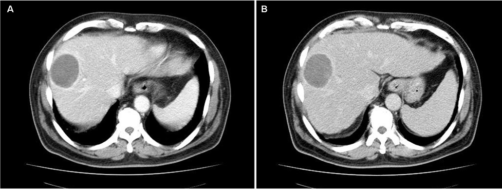
Fig. 1 Computed tomography scan shows ill-defined, 4×5 cm sized cystic mass located in segment V/VIII with peripheral enhancement in the arterial phase, panel (A) and venous phase, panel (B).
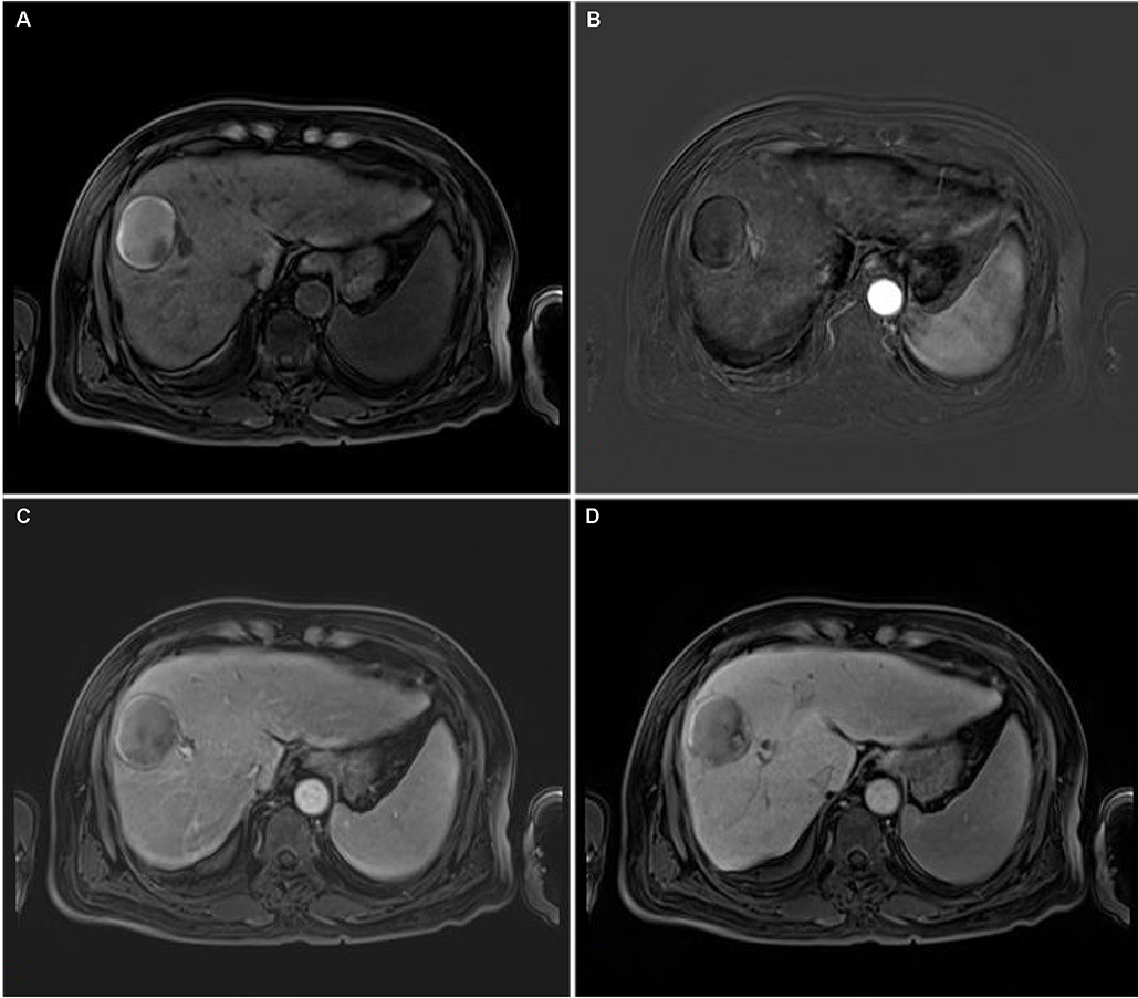
Fig. 2 Dynamic magnetic resonance imaging shows; pre-enhancement phase (A), arterial phase (B), portal phase (C), papillary-shaped projection from cyst wall toward the inner side at two-minute delay phase (D).
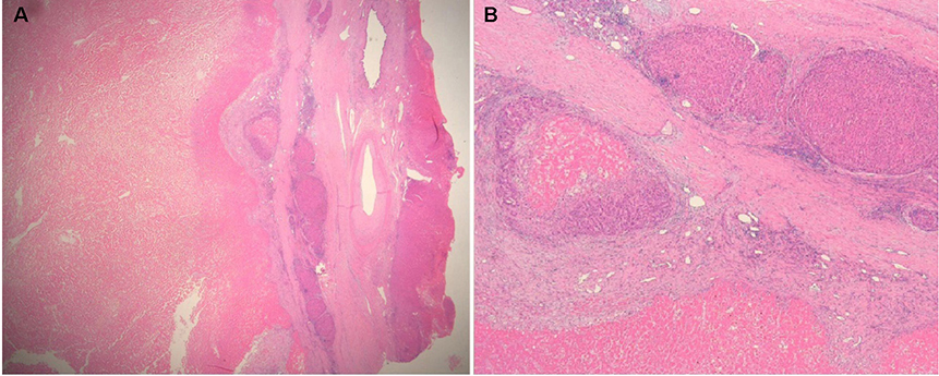
Fig. 3 Remnant hepatocellucar carcinoma cell surrounding broad necrosis (A, H&E, ×40), pleomorphic and undifferentiated malignant cell (B, H&E, ×200).
Reference
-
1. Gonwa ME, Casillas J, Livingstone AS, Robinson PG. Cystic hepatocellular carcinoma: CT findings. J Comput Assist Tomogr. 1991; 15:1045–1047.2. Yoo YJ, Kim JH. Spontaneous complete remission of hepatocellular carcinoma. Korean J Gastroenterol. 2015; 66:359–362.
Article3. Abiru S, Kato Y, Hamasaki K, Nakao K, Nakata K, Eguchi K. Spontaneous regression of hepatocellular carcinoma associated with elevated levels of interleukin 18. Am J Gastroenterol. 2002; 97:774–775.
Article4. Park HS, Jang KY, Kim YK, Cho BH, Moon WS. Hepatocellular carcinoma with massive lymphoid infiltration: a regressing phenomenon? Pathol Res Pract. 2009; 205:648–652.
Article5. Gottfried EB, Steller R, Paronetto F, Lieber CS. Spontaneous regression of hepatocellular carcinoma. Gastroenterology. 1982; 82:770–774.
Article6. Sun J, Yu X, Wang C, et al. RIP-1/c-FLIPL induce hepatic cancer cell apoptosis through regulating tumor necrosis factor-related apoptosis-inducing ligand (TRAIL). Med Sci Monit. 2017; 23:1190–1199.
Article7. Parks AL, McWhirter RM, Evason K, Kelley RK. Cases of spontaneous tumor regression in hepatobiliary cancers: implications for immunotherapy? J Gastrointest Cancer. 2015; 46:161–165.
Article8. Bruix J, Sherman M, American Association. Management of hepatocellular carcinoma: an update. Hepatology. 2011; 53:1020–1022.
Article9. Falidas E, Pazidis A, Anyfantakis G, Vlachos K, Goudeli C, Villias C. Multicystic hepatocarcinoma mimicking liver abscess. Case Rep Surg. 2013; 2013:374905.
Article10. Hagiwara S, Ogino T, Takahashi Y, et al. Hepatocellular carcinoma mimicking liver abscesses in a cirrhotic patient with severe septic shock as a result of salmonella O9 HG infection. Case Rep Gastroenterol. 2009; 3:56–60.
Article11. Choi BI, Lim JH, Han MC, et al. Biliary cystadenoma and cystadenocarcinoma: CT and sonographic findings. Radiology. 1989; 171:57–61.
Article12. Kim SH, Jeong WK, Kim Y, et al. Hepatocellular carcinoma composed of two different histologic types: imaging features on gadoxetic acid-enhanced liver MRI. Clin Mol Hepatol. 2013; 19:92–96.
Article
- Full Text Links
-
- Actions
-
Cited
- CITED
-
- Close
- Share
-
- Similar articles
-
- Duodenal Mucinous Carcinoma: A Case Report
- Cystic Hepatocellular carcinoma: a case report
- Fine Needle Aspiration Cytology of Mucinous Cystic Carcinoma of the Pancreas: A Case Report
- Differential diagnosis of cystic diseases of pancreas
- Pancreatic Collision Tumor of Desmoid-Type Fibromatosis and Mucinous Cystic Neoplasm: A Case Report
