Duodenal Mucinous Carcinoma: A Case Report
- Affiliations
-
- 1Department of Radiology, Dankook University Hospital, Dankook University College of Medicine, Cheonan, Korea. jkn1303@dreamwiz.com
- KMID: 2161171
- DOI: http://doi.org/10.3348/jksr.2015.72.3.185
Abstract
- Duodenal mucinous carcinoma is exceedingly rare and a case report about duodenal mucinous carcinoma in a 61-year-old man mimicking pancreatic cystic neoplasm by radiological evaluation, endoscopy, and even surgical findings is presented.
MeSH Terms
Figure
-
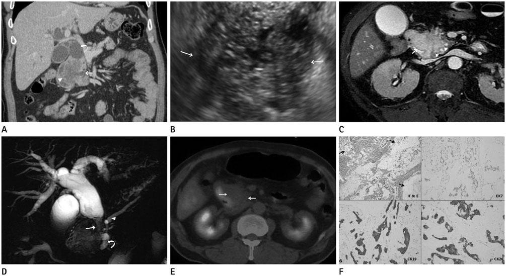
Fig. 1 Duodenal mucinous carcinoma with invasion of pancreas in a 61-year-old male patient. A. Contrast-enhanced CT image with coronal reformation shows a low-attenuating mass with partly irregular mucosal surface (arrowhead) in the duodenum, and dilatation of the pancreatic duct (arrow) and the bile duct (curved arrow). B. Endoscopic ultrasonography shows heterogeneous hyperechoic mass (arrows) with scattered anechoic spots. C. Axial fast imaging employing steady-state acquisition MR imaging shows a high signal-intensity cystic mass (arrow) bulging into the duodenal lumen and irregular tumor infiltration (arrowheads) around the distal common bile duct in the pancreatic head. D. MR cholangiopancreatography shows severe dilatation of the bile duct with segmental luminal narrowing (arrow) at the distal common bile duct and moderate dilatation of the main pancreatic duct (arrowhead) and branches (curved arrow) of pancreatic ducts. E. Fluorodeoxyglucose positron emission tomography-CT fusion imaging shows no increased uptake in the mass of pancreatic head (arrows). F. Photomicroscopic pathology (hematoxylin and eosin × 10, cytokeratin immunohistochemistry 7, 19, and 20) shows tumor cell infiltrations in the duodenal mucosa and mural layers with abundant extracellular mucin (arrows) and positive staining in cytokeratin 19 and 20 and negative in 7.
Reference
-
1. DiSario JA, Burt RW, Vargas H, McWhorter WP. Small bowel cancer: epidemiological and clinical characteristics from a population-based registry. Am J Gastroenterol. 1994; 89:699–701.2. Okumura F, Senoo K, Yoshida M, Miyabe K, Naito I, Tanaka H, et al. [A case of peritoneal dissemination from mucinous carcinoma of the duodenum, which was associated with tumor thrombosis in the accessory pancreatic duct and successfully treated by chemotherapy]. Nihon Shokakibyo Gakkai Zasshi. 2009; 106:1736–1743.3. Tsuro K, Matsumoto M, Moriyasu H, Nakatani Y, Sakurai S, Maekawa Y, et al. A case of duodenal mucinous adenocarcinoma infiltrating the cystic duct diagnosed as transpapillary with transnasal endoscopy. Dig Endosc. 2010; 22:246–247.4. Yagyu T, Aihara T, Murayama M, Kikuchi S, Nakamura E, Hase K, et al. Mucinous carcinoma of the duodenum associated with hereditary nonpolyposis colorectal cancer: report of a case. Surg Today. 2006; 36:1129–1132.5. Rosai J. Rosai and Ackerman's Surgical Pathology. In : Rosai J, editor. Gastrointestinal tract: esophagus, stomach, small bowel, appendix, large bowel, anus. 10th ed. Philadelphia: Elsevier-Mosby;2011. p. 731–755.6. Ko EY, Ha HK, Kim AY, Yoon KH, Yoo CS, Kim HC, et al. CT differentiation of mucinous and nonmucinous colorectal carcinoma. AJR Am J Roentgenol. 2007; 188:785–791.7. Hussain SM, Outwater EK, Siegelman ES. Mucinous versus nonmucinous rectal carcinomas: differentiation with MR imaging. Radiology. 1999; 213:79–85.8. Berger KL, Nicholson SA, Dehdashti F, Siegel BA. FDG PET evaluation of mucinous neoplasms: correlation of FDG uptake with histopathologic features. AJR Am J Roentgenol. 2000; 174:1005–1008.9. Itoh S, Ishiguchi T, Ishigaki T, Sakuma S, Maruyama K, Senda K. Mucin-producing pancreatic tumor: CT findings and histopathologic correlation. Radiology. 1992; 183:81–86.10. Kawamura H, Kondo Y, Osawa S, Nisida Y, Okada K, Isizu H, et al. A clinicopathologic study of mucinous adenocarcinoma of the stomach. Gastric Cancer. 2001; 4:83–86.
- Full Text Links
-
- Actions
-
Cited
- CITED
-
- Close
- Share
-
- Similar articles
-
- A Patient with Duodenal Mucinous Adenocarcinoma Presenting as a Laterally Spreading Tumor
- A Case of Cutaneous Mucinous Eccrine Carcinoma
- Ovarian Large Cell Neuroendocrine Carcinoma Associated with Endocervical-like Mucinous Borderline Tumor: A Case Report and Literature Review
- A Case of Cutaneous Mucinous Eccrine Carcinoma
- Mucocele-like Tumor of the Breast: A clinicopathologic case report
