Spontaneous Resolution of Iatrogenic Calcinosis Cutis after Parenteral Calcium Gluconate Therapy in Neonates
- Affiliations
-
- 1Department of Orthopedic Surgery, Keimyung University School of Medicine, Daegu, Korea. skspos@dsmc.or.kr
- KMID: 2444787
- DOI: http://doi.org/10.4055/jkoa.2019.54.2.192
Abstract
- Iatrogenic calcinosis cutis is due to the intravenous administration of calcium gluconate or calcium chloride to treat hypocalcemia. The arthors report three cases of calcinosis cutis with calcifications involving the upper or lower extremities in neonates following the extravasation of calcium gluconate. Three neonates, a 2-week-old girl, 4-week-old boy, and a 4-week-old girl, were consulted for indurated nodules after the intravenous administration of calcium gluconate at the intensive care unit. Complete remission of palpable nodule and calcification was observed on the radiograph at three weeks, four weeks and six months after the initial presentation in each. All three neonates with iatrogenic calcinosis curtis were resolved spontaneously without functional and cosmetic complications. According to enhancement of the patient's cognition about benign disease, a suitable explanation of the disease and avoiding unnecessary treatment through an early diagnosis of iatrogenic calcinosis cutis will reduce a number of potential medical malpractice disputes.
Keyword
MeSH Terms
Figure
-

Figure 1 (A) Anteroposterior and lateral radiographs from a 27-day-old boy shows scattered calcifications on the posteromedial area of the right ankle (arrows). (B) Smaller lesion size and more lucent density of calcifications one week after the initial presentation (arrows). (C) Four weeks later, calcification is completely absent.

Figure 2 (A) Radiographs from a 26-day-old girl, show a huge, diffuse, scattered calcification of the right forearm. (B) Smaller size and more lucent density of calcification three weeks later. (C) Six months after her initial presentation, the calcification disappeared spontaneously without any specific treatment.
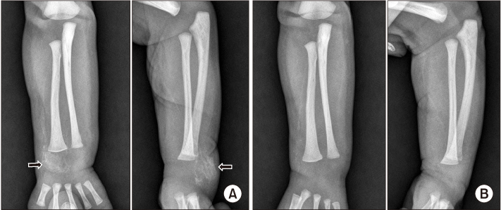
Figure 3 (A) Radiographs from a 14-day-old girl show a large, hazy calcification on the radial side of the right wrist (arrows). (B) Three weeks later, the calcification disappeared without specific treatment.
Reference
-
1. Caksen H, Odabaş D. An infant with gigantic subcutaneous calcium deposition following extravasation of calcium gluconate. Pediatr Dermatol. 2002; 19:277–279.2. Goldminz D, Barnhill R, McGuire J, Stenn KS. Calcinosis cutis following extravasation of calcium chloride. Arch Dermatol. 1988; 124:922–925.
Article3. Hironaga M, Fujigaki T, Tanaka S. Cutaneous calcinosis in a neonate following extravasation of calcium gluconate. J Am Acad Dermatol. 1982; 6:392–395.
Article4. Kagen MH, Bansal MG, Grossman M. Calcinosis cutis following the administration of intravenous calcium therapy. Cutis. 2000; 65:193–194.5. Jucglà A, Sais G, Curco N, Marcoval J, Moreno A, Peyri J. Calcinosis cutis following liver transplantation: a complication of intravenous calcium administration. Br J Dermatol. 1995; 132:275–278.6. Sahn EE, Smith DJ. Annular dystrophic calcinosis cutis in an infant. J Am Acad Dermatol. 1992; 26:1015–1017.
Article7. Rodríguez-Cano L, García-Patos V, Creus M, Bastida P, Ortega JJ, Castells A. Childhood calcinosis cutis. Pediatr Dermatol. 1996; 13:114–117.
Article8. Moss J, Syrengelas A, Antaya R, Lazova R. Calcinosis cutis: a complication of intravenous administration of calcium glucanate. J Cutan Pathol. 2006; 33:Suppl 2. 60–62.
Article9. Duhn R, Schoen EJ, Siu M. Subcutaneous fat necrosis with extensive calcification after hypothermia in two newborn infants. Pediatrics. 1968; 41:661–664.
Article10. Lee FA, Gwinn JL. Roentgen patterns of extravasation of calcium gluconate in the tissues of the neonate. J Pediatr. J Pediatr. 1975; 86:598–601.
- Full Text Links
-
- Actions
-
Cited
- CITED
-
- Close
- Share
-
- Similar articles
-
- Two Cases of Iatrogenic Calcinosis Cutis Following Extravasation of Calcium Gluconate in Neonates
- Unusual Presentation of Calcinosis Cutis: Venous Calcification Following intravenous Calcium Gluconate Administration in a Preterm Baby
- A Case of Calcinosis Cutis due to Intravenous Administration of Calcium Gluconate
- An Unusual Case of Transepidermal Elimination of Calcinosis Cutis
- A rare case report of neonatal calcinosis cutis induced by distant and delayed extravasation of intravenous calcium gluconate
