Granulocytic Sarcoma of the Sciatic Nerve Manifested as Relapse of Acute Myeloid Leukemia: A Case Report
- Affiliations
-
- 1Department of Radiology, 3Orthopedic Surgery, Yeouido St. Mary's Hospital, The Catholic University of Korea College of Medicine, Seoul, Korea. jmpark@catholic.ac.kr
- 2Department of Hospital Pathology, Yeouido St. Mary's Hospital, The Catholic University of Korea College of Medicine, Seoul, Korea.
- 3Department ofOrthopedic Surgery, Yeouido St. Mary's Hospital, The Catholic University of Korea College of Medicine, Seoul, Korea.
- KMID: 2442524
- DOI: http://doi.org/10.3348/jksr.2019.80.2.339
Abstract
- Granulocytic sarcoma is a form of extramedullary involvement of primitive myeloid cells. A 69-year-old male patient, with history of acute myeloid leukemia (AML) in remission state for 4 years, presented numbness, radiating pain and progressive motor weakness in left leg. MRI showed perineural thickening of the left sciatic nerve with increased signal intensity on fat-saturated T2-weighted image. The patient underwent surgical excision and the pathology was confirmed as granulocytic sarcoma. Its involvement of the peripheral nerve is extremely rare and also unusual to be the only evidence of AML relapse. In this case, we figured out the MRI feature of granulocytic sarcoma involving sciatic nerve, emerged as a sole manifestation of AML relapse.
MeSH Terms
Figure
-
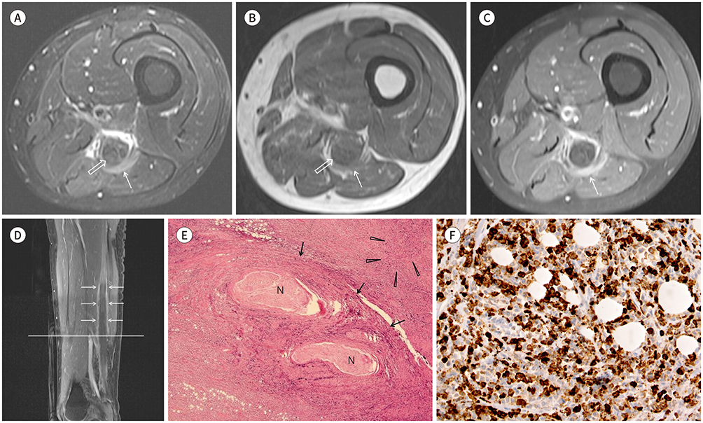
Fig. 1 A 69-year-old-man with granulocytic sarcoma in sciatic nerve. A, B. Axial fat-saturated T2-weighted image (A) and axial T1-weighted image (B) of MRI of left thigh show diffuse swelling of left sciatic nerve with intermediate T2 and T1 signal intensity (open arrows) and irregular peripheral nerve sheath thickening with heterogenous hyperintense T2 signal intensity and isointense T1 signal intensity (thin arrows) at distal thigh level. C. Contrast-enhanced T1-weighted image shows thick and circumferential enhancement of peripheral nerve sheath (arrow) with internal mild, mottled enhancement of nerve fascicles. D. Sagittal contrast-enhanced T1-weighted image shows an about 19 cm length of diffuse fusiform thickening and intense enhancement of left sciatic nerve sheath at mid to distal thigh level (arrows). The line indicates the transverse section level of Fig. 1A–C. E. Histopathology of epineurium and surrounding soft tissue in surgical specimen reveals diffuse infiltration of atypical tumor cells around the nerve fibers and surrounding soft tissue (hematoxylin and eosin stain, × 12.5). Atypical tumor cells are widely infiltrating epineurium (arrows) and surrounding soft tissue (arrowheads). Specimen contains two small nerve bundles (N) that passes through epineurium. Main nerve fascicle is not included in this specimen. F. The immunohistochemical staining of tumor cells shows diffuse positive for myeloperoxidase. This result demonstrates that the tumor cells are leukemic cells derived from the myeloid lineage and is important in the diagnosis of granulocytic sarcoma (myeloperoxidase stain, × 400).
Reference
-
1. Yilmaz AF, Saydam G, Sahin F, Baran Y. Granulocytic sarcoma: a systematic review. Am J Blood Res. 2013; 3:265–270.2. Seok JH, Park J, Kim SK, Choi JE, Kim CC. Granulocytic sarcoma of the spine: MRI and clinical review. AJR Am J Roentgenol. 2010; 194:485–489.
Article3. Novick SL, Nicol TL, Fishman EK. Granulocytic sarcoma (chloroma) of the sacrum: initial manifestation of leukemia. Skeletal Radiol. 1998; 27:112–114.
Article4. Lee KH, Lee JH, Choi SJ, Lee JH, Kim S, Seol M, et al. Bone marrow vs extramedullary relapse of acute leukemia after allogeneic hematopoietic cell transplantation: risk factors and clinical course. Bone Marrow Transplant. 2003; 32:835–842.
Article5. Clark WB, Strickland SA, Barrett AJ, Savani BN. Extramedullary relapses after allogeneic stem cell transplantation for acute myeloid leukemia and myelodysplastic syndrome. Haematologica. 2010; 95:860–863.
Article6. Arrigan M, Smyth L, Harmon M, Flynn C, Sheehy N. Imaging findings in recurrent extramedullary leukaemias. Cancer Imaging. 2013; 13:26–35.
Article7. Stillman MJ, Christensen W, Payne R, Foley KM. Leukemic relapse presenting as sciatic nerve involvement by chloroma (granulocytic sarcoma). Cancer. 1988; 62:2047–2050.
Article8. Liu HC, Hung GY, Yen HJ, Hsieh MY, Chiou TJ. Acute sciatica: an unusual presentation of extramedullary relapse of acute lymphoblastic leukemia. Int J Hematol. 2007; 86:163–165.
Article9. Warme B, Sullivan J, Tigrani DY, Fred DM. Chloroma of the forearm: a case report of leukemia recurrence presenting with compression neuropathy and tenosynovitis. Iowa Orthop J. 2009; 29:114–116.10. Ooi GC, Chim CS, Khong PL, Au WY, Lie AK, Tsang KW, et al. Radiologic manifestations of granulocytic sarcoma in adult leukemia. AJR Am J Roentgenol. 2001; 176:1427–1431.
Article
- Full Text Links
-
- Actions
-
Cited
- CITED
-
- Close
- Share
-
- Similar articles
-
- Granulocytic Sarcoma in the Leg Mimicking Hemorrhagic Abscess
- A case of isolated granulocytic sarcoma of the kidney after complete remission of acute myelocytic leukemia
- A Case of Temporal Bone Myeloid Sarcoma
- A case of granulocytic sarcoma of the pancreas and kidney in a patient presenting with jaundice
- A Case of Granulocytic Sarcoma Involving the Forniceal Conjunctiva
