Metastasis of Rhabdomyosarcoma to the Male Breast: a Case Report with Magnetic Resonance Imaging Findings
- Affiliations
-
- 1Department of Radiology, St. Vincent's Hospital, The Catholic University of Korea, Suwon, Korea.
- 2Department of Radiology, Seoul St. Mary's Hospital, The Catholic University of Korea, Seoul, Korea. gmlionmain@gmail.com
- 3Department of Pathology, Seoul St. Mary's Hospital, The Catholic University of Korea, Seoul, Korea.
- KMID: 2442237
- DOI: http://doi.org/10.13104/imri.2019.23.1.75
Abstract
- Metastasis of rhabdomysarcoma to the breast is a very rare manifestation in adult males. Herein, we report a case of metastasis from embryonal rhabdomyosarcoma in the left hypothenar muscle that presented as a breast mass in a 38-year-old man, who four months later expired because of multiple bone metastases related to pancytopenia. We describe the various imaging findings, including mammograms, ultrasonography, computerized tomography (CT), positron emission tomography-computed tomography (PET-CT), and magnetic resonance imaging (MRI) of this rare disease. The various imaging findings of this lesion could be helpful for future diagnosis of male breast lesions.
Keyword
MeSH Terms
Figure
-
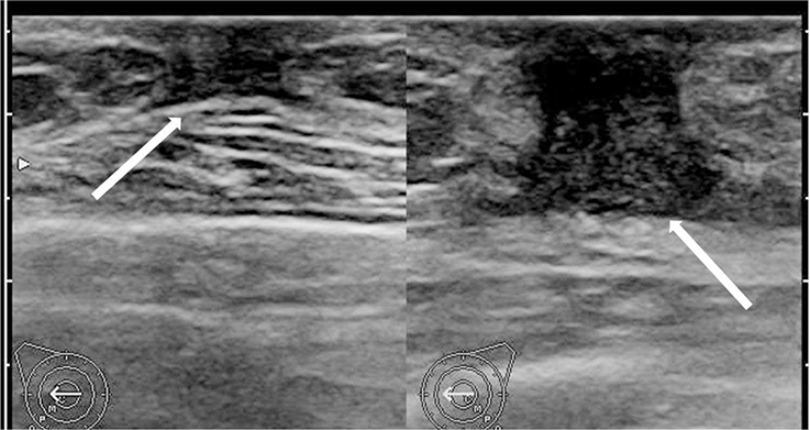
Fig. 1 Ultrasonographic image reveals hypoechoic fibroglandular tissue in both subareolar spaces, more on the left, suggesting gynecomastia (arrows).
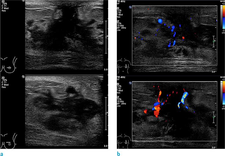
Fig. 2 Follow-up ultrasonographic image (a) and color Doppler scan (b) reveals an irregular mass of about 4 × 1.7 cm with a heterogeneous internal echo pattern in the left subareolar-to-central region with internal vascularity, suggesting metastasis of a known rhabdomyosarcoma.

Fig. 3 Mammograms of the left breast reveal an irregular, circumscribed, hyperdense mass of about 5.9 cm in the left subareolar-to-central region.
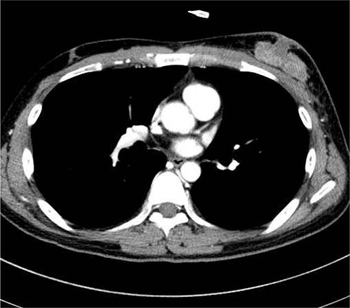
Fig. 4 Contrast-enhanced CT image reveals a lobulated, heterogeneous enhancing mass of about 5.3 cm in the left subareolar-to-central region.
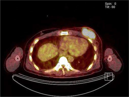
Fig. 5 Positron-emission tomography-computed tomography (PET-CT) shows a hypermetabolic mass in the left breast (SUVmax: 11.8).

Fig. 6 T1- (a), T2- (b), fat-suppressed T2- (c), and 2 minute delayed gadolinium-enhanced T1-weighted (d) MR images. An axial MRI showed isointense signal intensity, compared to the muscle in contrast T1 weighted image (WI), with high SI on T2WI. The signal of the mass was not suppressed on fat-suppressed T2WI and showed heterogeneous enhancement on contrast-enhanced T1WI with high SI on a high b value diffusion-weighted image (b = 800 s/mm2) (e), with a definitive low apparent diffusion coefficient (mean 0.69 × 10⁻³s/mm2) (f) in the left breast.
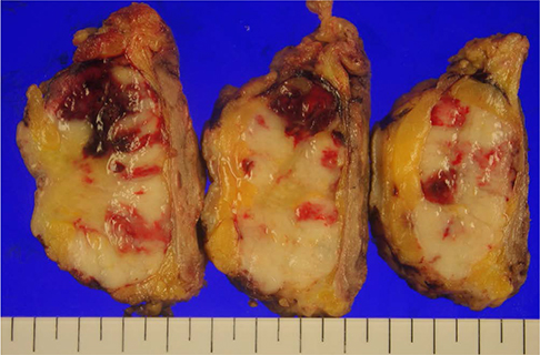
Fig. 7 A well-demarcated tumor mass, 5.0 × 5.0 × 3.0 cm, pale brown, solid cut surfaces with hemorrhagic foci.
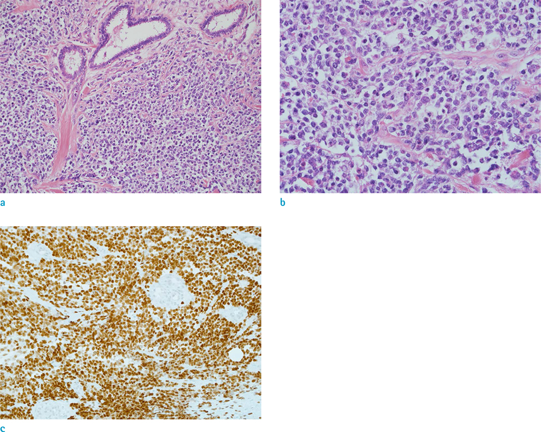
Fig. 8 Metastatic rhabdomyosarcoma was confirmed by pathology. (a) Tumor cells are small, round to oval cells, diffusely infiltrating the breast parenchyma (× 200, Hematoxylin & Eosin [H&E] staining). (b) Tumor cells show eccentrically located vesicular nuclei and clear-to-eosinophilic cytoplasm (× 400, H&E staining). (c) Tumor cells are diffusely positive for MyoD1 (× 200 MyoD1 immunohistochemical staining).
Reference
-
1. Ferrari A, Dileo P, Casanova M, et al. Rhabdomyosarcoma in adults. A retrospective analysis of 171 patients treated at a single institution. Cancer. 2003; 98:571–580.2. Birjawi GA, Haddad MC, Tawil AN, Khoury NJ. Metastatic rhabdomyosarcoma to the breast. Eur Radiol. 2001; 11:555–558.3. Hays DM, Donaldson SS, Shimada H, et al. Primary and metastatic rhabdomyosarcoma in the breast: neoplasms of adolescent females, a report from the Intergroup Rhabdomyosarcoma Study. Med Pediatr Oncol. 1997; 29:181–189.
Article4. Kebudi R, Koc BS, Gorgun O, Celik A, Kebudi A, Darendeliler E. Breast metastases in children and adolescents with rhabdomyosarcoma: a large single-institution experience and literature review. J Pediatr Hematol Oncol. 2017; 39:67–71.
Article5. Noguera J, Martinez-Miravete P, Idoate F, et al. Metastases to the breast: a review of 33 cases. Australas Radiol. 2007; 51:133–138.
Article6. Surov A, Fiedler E, Holzhausen HJ, Ruschke K, Schmoll HJ, Spielmann RP. Metastases to the breast from nonmammary malignancies: primary tumors, prevalence, clinical signs, and radiological features. Acad Radiol. 2011; 18:565–574.7. Toombs BD, Kalisher L. Metastatic disease to the breast: clinical, pathologic, and radiographic features. AJR Am J Roentgenol. 1977; 129:673–676.
Article8. Nguyen HV, Aminololama-Shakeri S, Zhang Y. Initial presentation and recurrence of metastatic rhabdomyosarcoma as breast mass. Radiol Case Rep. 2013; 8:855.
Article9. Howarth CB, Caces JN, Pratt CB. Breast metastases in children with rhabdomyosarcoma. Cancer. 1980; 46:2520–2524.
Article
- Full Text Links
-
- Actions
-
Cited
- CITED
-
- Close
- Share
-
- Similar articles
-
- Giant Breast Involvement in Acute Lymphoblastic Leukemia: MRI Findings
- An Unusual Imaging Finding of Breast Metastasis from Rhabdomyosarcoma
- The CT and MR Imaging Findings of Adulthood Sinonasal Alveolar Rhabdomyosarcoma with Disseminated Metastases on 18F-FDG PET/CT: Report of Two Cases
- A Rare Case of Male Primary Breast Lymphoma
- Sinonasal Rhabdomyosarcoma Metastasis in Bilateral Multiple Extraocular Muscles: A Case Report and Brief Literature Review
