The CT and MR Imaging Findings of Adulthood Sinonasal Alveolar Rhabdomyosarcoma with Disseminated Metastases on 18F-FDG PET/CT: Report of Two Cases
- Affiliations
-
- 1Department of Radiology, Dongsan Medical Center, Keimyung University School of Medicine, Daegu, Korea. sklee@dsmc.or.kr
- KMID: 2097926
- DOI: http://doi.org/10.3348/jksr.2011.64.2.117
Abstract
- We report herein two cases of adulthood sinonasal alveolar rhabdomyosarcoma that showed intense hypermetabolism and disseminated metastasis on 18F-FDG PET/CT. The contrast-enhanced CT (CECT) and gadolinium-enhanced T1-weighted images (Gd-T1WI) showed multiple rings of intense enhancement (the "botryoid sign") of the mass. Adulthood rhabdomyosarcoma should be considered when a sinonasal mass shows the "botryoid sign" on CECT or Gd-T1WI, and intense hypermetabolism with disseminated metastases on 18F-FDG PET/CT.
MeSH Terms
Figure
-
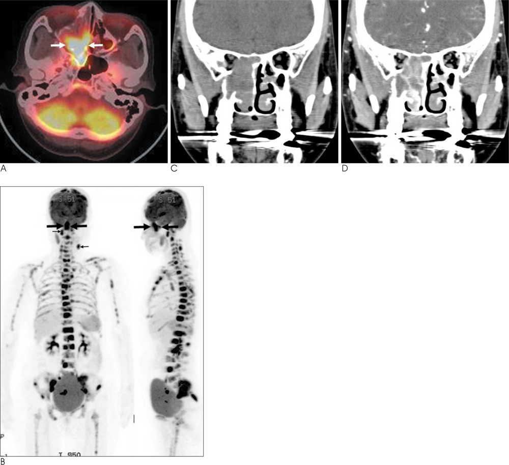
Fig. 1 Sinonasal rhabdomyosarcoma of the alveolar subtype with disseminated metastases to the bone marrow and lymph nodes in a 68-year-old woman. A. An axial 18F-FDG PET/CT image shows a lobular, intensely hypermetabolic mass (SUVmax = 14.6 g/mL) in the right sinonasal area (arrows). B. The coronal and sagittal maximum intensity projection images of 18F-FDG PET demonstrate intense hypermetabolism involving the axial and appendicular skeleton and the lymph nodes of the right retropharyngeal chain and the left level VA (small arrows), as well as the primary site (arrows). C. A coronal reformatted non-enhanced CT image shows a right-sided sinonasal mass with bony destruction. D. A coronal reformatted contrast-enhanced CT image reveals a mass with multiple rings of intense enhancement (the "botryoid sign").
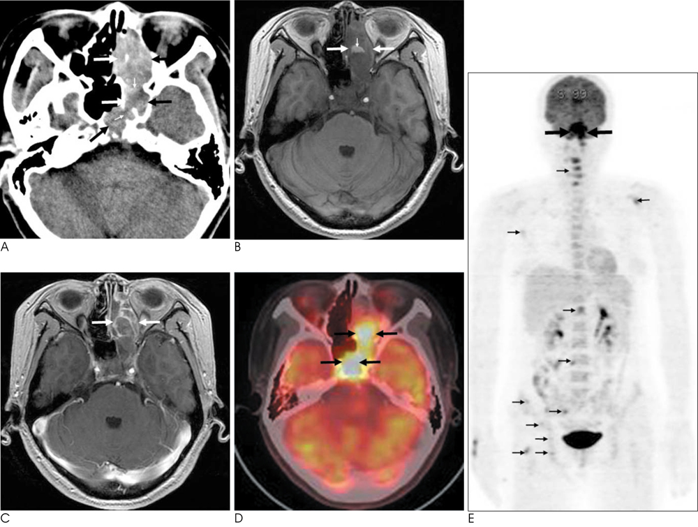
Fig. 2 Sinonasal rhabdomyosarcoma of the alveolar subtype with disseminated bone marrow and breast metastases in a 48-year-old woman. A. An axial non-enhanced CT image shows a large hyperattenuating mass (arrows) in the left nasal cavity, the ethmoid sinus and the bilateral sphenoid sinuses with bony destruction (small arrows). B. An axial spoiled gradient-echo T1-weighted image (T1WI) (TR/TE=7.3/3.0, FA=20) reveals a sinonasal mass (arrows), which is hypointense compared to that of the adjacent muscles, and the mass contains a focus of hyperintense hemorrhage (small arrow). C. An axial gadolinium-enhanced spoiled gradient-echo T1WI (TR/TE=7.3/3.0, FA=20) demonstrates a mass with multiple rings of intense enhancement (the "botryoid sign") (arrows). D. An axial 18F-FDG PET/CT image shows a lobular intensely hypermetabolic mass (SUVmax = 11.9 g/mL) in the sinonasal area (arrows). E. A coronal maximum intensity projection image of 18F-FDG PET demonstrates multiple bone marrow metastases involving the axial and appendicular skeleton (small arrows), as well as the primary site (arrows).
Reference
-
1. Feldman BA. Rhabdomyosarcoma of the head and neck. Laryngoscope. 1982; 92:424–440.2. La Quaglia MP, Heller G, Ghavimi F, Casper ES, Vlamis V, Hajdu S, et al. The effect of age at diagnosis on outcome in rhabdomyosarcoma. Cancer. 1994; 73:109–117.3. Tateishi U, Hosono A, Makimoto A, Nakamoto Y, Kaneta T, Fukuda H, et al. Comparative study of FDG PET/CT and conventional imaging in the staging of rhabdomyosarcoma. Ann Nucl Med. 2009; 23:155–161.4. Klem ML, Grewal RK, Wexler LH, Schöder H, Meyers PA, Wolden SL. PET for staging in rhabdomyosarcoma: an evaluation of PET as an adjunct to current staging tools. J Pediatr Hematol Oncol. 2007; 29:9–14.5. Peng F, Rabkin G, Muzik O. Use of 2-deoxy-2-[F-18]-fluoro-D-glucose positron emission tomography to monitor therapeutic response by rhabdomyosarcoma in children: report of a retrospective case study. Clin Nucl Med. 2006; 31:394–397.6. Iagaru A, Goris ML. Rhabdomyosarcoma diffusely metastatic to the bone marrow: suspicious findings on 99mTc-MDP bone scintigraphy confirmed by (18)F-18 FDG PET/CT and bone marrow biopsy. Eur J Nucl Med Mol Imaging. 2008; 35:1746.7. Seshadri N, Wright P, Balan KK. Rhabdomyosarcoma with widespread bone marrow infiltration: beneficial management role of F-18 FDG PET. Clin Nucl Med. 2007; 32:787–789.8. Hagiwara A, Inoue Y, Nakayama T, Yamato K, Nemoto Y, Shakudo M, et al. The "botryoid sign": a characteristic feature of rhabdomyosarcomas in the head and neck. Neuroradiology. 2001; 43:331–335.9. Enzinger FM, Weiss SW. Soft tissue tumors. 3rd ed. St Louis: Mosby;1995. p. 539–577.10. Ihara T, Okamura D, Takahashi N, Kohri M, Kayano H, Tamaru J, et al. Alveolar rhabdomyosarcoma mimicking nasal lymphoma at the initial presentation. J Clin Exp Hematop. 2008; 48:61–64.
- Full Text Links
-
- Actions
-
Cited
- CITED
-
- Close
- Share
-
- Similar articles
-
- Use of 18F-FDG PET/CT in Second Primary Cancer
- Metastases to Skeletal Muscles from Non-Small Cell Lung Cancer Demonstrated by 18F-FDG PET/CT
- Lung Adenocarcinoma Staged as an Unknown Primary Presenting with Symptomatic Colon Metastases: Staging by 18F-FDG PET/CT
- Orbital Metastases as Presenting Sign of Lung Carcinoma: Detection of Primary Malignancy and Disease Burden by F-18 FDG PET/CT
- Esophageal Leiomyoma with intense FDG uptake on 18F-FDG PET/CT
