
Giant Breast Involvement in Acute Lymphoblastic Leukemia: MRI Findings
- Affiliations
-
- 1Department of Radiology, Celal Bayar University of Medicine, Manisa, Turkey. slbasara@hotmail.com
- KMID: 2242205
- DOI: http://doi.org/10.4048/jbc.2012.15.2.258
Abstract
- Breast metastases in cases of leukemia are rare. We aimed to report the conventional-advanced magnetic resonance imaging (MRI) findings of unilateral breast involvement of acute lymphoblastic leukemia (ALL) and review the literature. A 32-year-old woman was first diagnosed with ALL in treated in 2004. She did not continue the follow-up after 2008. She was presented with a giant, progressive right breast palpable mass in 2010. Mass, contralateral breast tissue were evaluated with MRI, diffusion weighted imaging and MR spectroscopy. With MRI findings, lesion was evaluated as malignant, tru-cut biopsy revealed recurrence of ALL. Lymphoma, malignant melanoma, rhabdomyosarcoma are most common tumors metastase to breast. Breast metastases of leukemia are rare and occur primarily in patients with acute myeloid leukemia. Secondary ALL breast involvement is uncommon. In a patient with malignancy, any enlarging breast mass, even with benign radiologic appearance, should be investigated carefully and metastasis should not be forgotten.
MeSH Terms
Figure
-
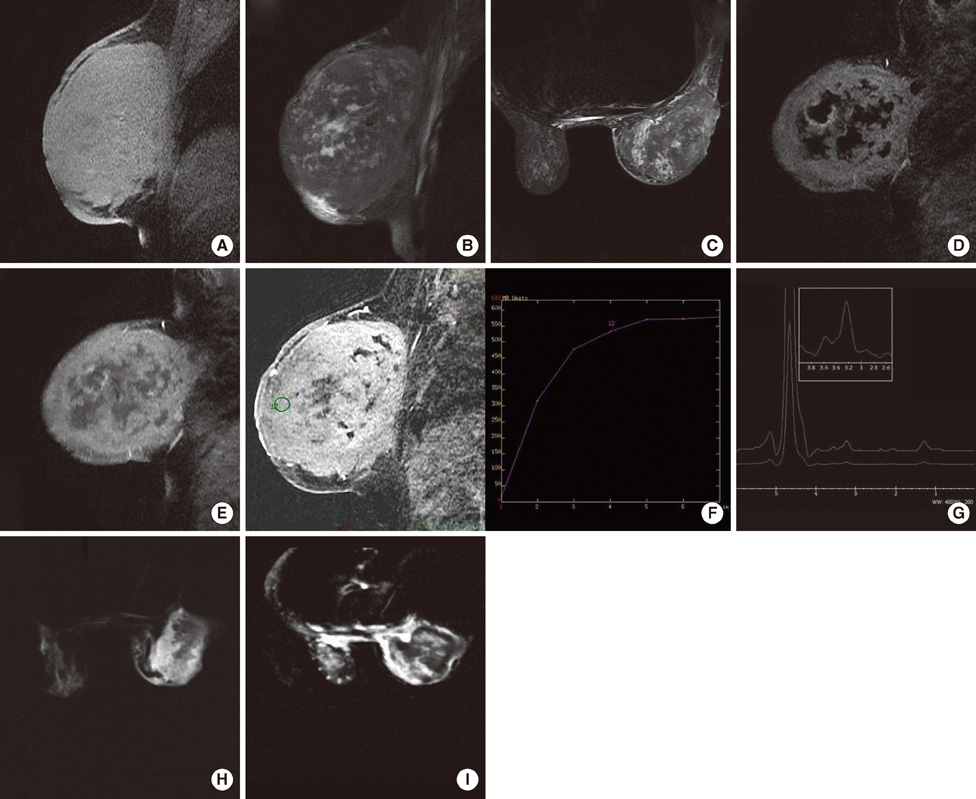
Figure 1 Acute lymphoblastic leukemia presenting as a giant breast mass in a 32-year-old woman. (A) Both the solid and necrotic components of the giant lesion in the right breast are hyperintense-isointense on precontrast fat saturated T1W images. (B) On fast spin echo, fat saturated T2W image of the solid and the necrotic parts of the lesion are hypointense-hyperintense. (C) On short TI inversion recovery image the signal characteristics are similar to the fat saturated T2W image. (D) Subtracted and (E) post contrast fat saturated T1W images, there is prominent enhancement. (F) Type 2 (plateau type, doubtful type) curve pattern is obtained from the enhanced involved breast tissue. (G) On diffusion weighted imaging (DWI), nearly the whole breast is hyperintense. (H) In the apparent coefficient diffusion (ADC) map, hyperintense parts on DWI image are hypointense indicating restricted diffusion. The necrotic parts are hyperintense. ADC value: 3.12×10⁻⁶ mm2/sec. (I) There is a prominent choline peak at 3.2 ppm in the BREASE sequence.
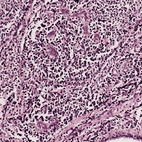
Figure 2 There is leukemic blasts infiltrating the breast tissue (H&E stain, ×100).
Cited by 2 articles
-
Granulocytic Sarcoma in Breast after Bone Marrow Transplantation
Seung Jin Kim, Woo Sung Hong, Sung Hyun Jun, Seong Hyun Jeong, Seok Youn Kang, Tae Hee Kim, Doo Kyoung Kang, Hyun Ee Yim, Yong Sik Jung, Ku Sang Kim
J Breast Cancer. 2013;16(1):112-116. doi: 10.4048/jbc.2013.16.1.112.Relapse of Biphenotypic Acute Leukemia as a Breast Mass
Hee-Chul Shin
J Breast Cancer. 2016;19(4):455-458. doi: 10.4048/jbc.2016.19.4.455.
Reference
-
1. Likaki-Karatza E, Mpadra FA, Karamouzis MV, Ravazoula P, Koukouras D, Margariti S, et al. Acute lymphoblastic leukemia relapse in the breast diagnosed with gray-scale and color Doppler sonography. J Clin Ultrasound. 2002. 30:552–556.
Article2. Khoury NJ, Hanna Al-Kass FM, Jaafar HN, Taher AT, Shamseddine AI. Bilateral breast involvement in acute myelogenous leukemia. Eur Radiol. 2000. 10:1031.
Article3. McCrea ES, Johnston C, Haney PJ. Metastases to the breast. AJR Am J Roentgenol. 1983. 141:685–690.
Article4. Hays DM, Donaldson SS, Shimada H, Crist WM, Newton WA Jr, Andrassy RJ, et al. Primary and metastatic rhabdomyosarcoma in the breast: neoplasms of adolescent females, a report from the Intergroup Rhabdomyosarcoma Study. Med Pediatr Oncol. 1997. 29:181–189.
Article5. Cunningham I. A clinical review of breast involvement in acute leukemia. Leuk Lymphoma. 2006. 47:2517–2526.
Article6. Yang WT, Muttarak M, Ho LW. Nonmammary malignancies of the breast: ultrasound, CT, and MRI. Semin Ultrasound CT MR. 2000. 21:375–394.
Article7. Yang WT, Metreweli C. Sonography of nonmammary malignancies of the breast. AJR Am J Roentgenol. 1999. 172:343–348.
Article8. Jakovljević B, Stevanović O, Bacić G. Metastases to the breast from small-cell lung cancer: MR findings. A case report. Acta Radiol. 2003. 44:485–488.
Article9. Koh DM, Collins DJ. Diffusion-weighted MRI in the body: applications and challenges in oncology. AJR Am J Roentgenol. 2007. 188:1622–1635.
Article10. Mountford C, Lean C, Malycha P, Russell P. Proton spectroscopy provides accurate pathology on biopsy and in vivo. J Magn Reson Imaging. 2006. 24:459–477.
Article11. West KW, Rescorla FJ, Scherer LR 3rd, Grosfeld JL. Diagnosis and treatment of symptomatic breast masses in the pediatric population. J Pediatr Surg. 1995. 30:182–186.
Article12. Rogers DA, Lobe TE, Rao BN, Fleming ID, Schropp KP, Pratt AS, et al. Breast malignancy in children. J Pediatr Surg. 1994. 29:48–51.
Article
- Full Text Links
-
- Actions
-
Cited
- CITED
-
- Close
- Share
-
- Similar articles
-
- Acute Lymphoblastic Leukemia Presented as Multiple Breast Masses
- Precursor B-Cell Acute Lymphoblastic Leukemia in Two Patients with a History of Cytotoxic Therapy
- A case of acute lymphoblastic leukemia complicating neuroblastoma in remission
- Unusual isolated extramedullary relapse of acute lymphoblastic leukemia in the breast despite complete donor hematopoietic chimerism after allogeneic hematopoietic stem cell transplantation
- A case of bone marrow necrosis in acute lymphoblastic leukemia


