Ann Dermatol.
2019 Apr;31(2):213-216. 10.5021/ad.2019.31.2.213.
Congenital Dermal Melanocytosis on the Foot: A Case Report and Review of the Literature
- Affiliations
-
- 1Department of Dermatology, Seoul National University College of Medicine, Seoul, Korea. jehomun@gmail.com
- 2Department of Pathology, Seoul National University College of Medicine, Seoul, Korea.
- 3Institute of Human-Environment Interface Biology, Seoul National University, Seoul, Korea.
- KMID: 2439069
- DOI: http://doi.org/10.5021/ad.2019.31.2.213
Abstract
- Dermal melanocytosis is a common pigmented skin disease, characterized by an increased number of ectopic melanocytes in the dermis. Rare variants of dermal melanocytosis that do not belong to these four typical groups-nevus of Ota, nevus of Ito, blue nevus, and Mongolian spots-are called dermal melanocyte hamartoma, or congenital dermal melanocytosis (CDM) as it mostly appears from birth. We report a case of CDM on the foot of a young woman with a literature review of previously reported cases of CDM.
Keyword
MeSH Terms
Figure
-
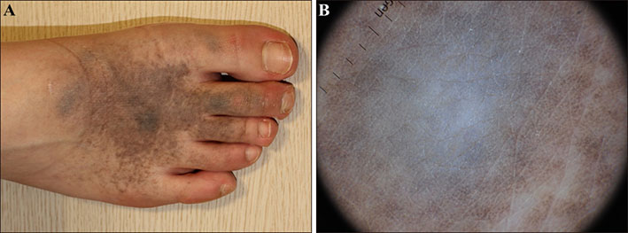
Fig. 1 (A) Mottled, confluent, blue-gray macules surrounding darker patches on the right dorsum of the foot. (B) Steel blue or gray structureless area surrounded by mottled brown globules on dermoscopy.

Fig. 2 (A~C) Increased and scattered pigmented melanocytes without nest formation throughout the dermis. Scattered and elongated melanocytes among collagen bundles without certain orientation (H&E; A: ×100, B: ×200, C: ×400). (D) Melanocytes were positively stained for Melan-A (MART-1, ×200).
Reference
-
1. Lee S, Kim DH, Lee G, Whang KU, Lee JS, Park YL. An unusual case of congenital dermal melanocytosis. Ann Dermatol. 2010; 22:460–462.
Article2. Burkhart CG, Gohara A. Dermal melanocyte hamartoma. A distinctive new form of dermal melanocytosis. Arch Dermatol. 1981; 117:102–104.
Article3. Kim S, Kim JA, Kim WS, Lee JH, Yang JM. Congenital dermal melanocytosis confined to the palm. J Eur Acad Dermatol Venereol. 2007; 21:1116–1117.
Article4. Pessach Y, Goldberg I, Sprecher E, Gat A, Harel A. An unusual presentation of congenital dermal melanocytosis fitting the rare diagnosis of dermal melanocyte hamartoma. Cutis. 2014; 94:E16–E17.5. Franceschini D, Dinulos JG. Dermal melanocytosis and associated disorders. Curr Opin Pediatr. 2015; 27:480–485.
Article6. Bashiti HM, Blair JD, Triska RA, Keller L. Generalized dermal melanocytosis. Arch Dermatol. 1981; 117:791–793.
Article7. Vélez A, Fuente C, Belinchón I, Martín N, Furió V, Sánchez Yus E. Congenital segmental dermal melanocytosis in an adult. Arch Dermatol. 1992; 128:521–525.
Article8. Grézard P, Berard F, Balme B, Perrot H. Congenital bilateral dermal melanocytosis with a dermatomal pattern. Dermatology. 1999; 198:105–106.
Article9. Krishnan RS, Roark TR, Hsu S. Isolated patch of speckled, congenital, pigmented dermal melanocytosis outside the face or acromioclavicular regions. J Eur Acad Dermatol Venereol. 2003; 17:238–239.
Article10. Kim S, Park JH, Kim JA, Lee JH, Yang JM, Lee ES, et al. Congenital combined dermal and epidermal melanocytosis: a new entity? J Eur Acad Dermatol Venereol. 2007; 21:1282–1283.
Article
- Full Text Links
-
- Actions
-
Cited
- CITED
-
- Close
- Share
-
- Similar articles
-
- A Case of Acquired Dermal Melanocytosis Occurring on the Chest
- An Unusual Case of Congenital Dermal Melanocytosis
- Acquired Dermal Melanocytosis Occurring on the Hand
- A Case of Uncommon Acquired Dermal Melanocytosis
- Two Cases of Vitiligo Developed on the Persisting Dermal Melanocytosis: Is There a Difference between Epidermal Melanocytes and Dermal Melanocytes?
