Ann Dermatol.
2010 Nov;22(4):460-462. 10.5021/ad.2010.22.4.460.
An Unusual Case of Congenital Dermal Melanocytosis
- Affiliations
-
- 1Department of Dermatology, College of Medicine, Soonchunhyang University, Seoul, Korea. ylpark@schmc.ac.kr
- KMID: 2266188
- DOI: http://doi.org/10.5021/ad.2010.22.4.460
Abstract
- Dermal melanocytosis is characterized by the presence of ectopic melanocytes in the dermis. The most common forms include the Mongolian spot, blue nevus, nevus of Ota, and nevus of Ito. Some types of dermal melanocytosis do not fit into any of these morphologic categories, however. Our case demonstrated an extensive amount of uniform deep blue patches of nevi with unilateral distribution on the left face, neck, chest, shoulder, and back. On histopathologic examination, a number of elongated melanocytes scattered throughout the dermis were found. We herein report a case of congenital unilateral dermal melanocytosis.
Figure
-
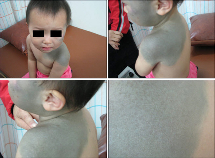
Fig. 1 An extensive amount of uniform deep blue patches were seen to cover the left unilateral side of the face, neck, chest, shoulder, and back.
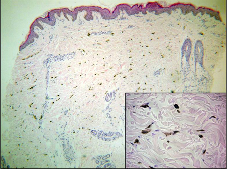
Fig. 2 Melanocytes were numerous in the upper and middle dermis. Melanocytes were scattered among the collagen bundles, and some of these cells were aggregated around the blood vessels. Their elongated cytoplasms were loaded with fine melanin. Melanophages were not seen (H&E, ×40, inset ×400).
Cited by 1 articles
-
Congenital Dermal Melanocytosis on the Foot: A Case Report and Review of the Literature
Soo Ick Cho, Jungyoon Moon, Gwanghyun Jo, Cheol Lee, Je-Ho Mun
Ann Dermatol. 2019;31(2):213-216. doi: 10.5021/ad.2019.31.2.213.
Reference
-
1. Bashiti HM, Blair JD, Triska RA, Keller L. Generalized dermal melanocytosis. Arch Dermatol. 1981. 117:791–793.
Article2. Burkhart CG, Gohara A. Dermal melanocyte hamartoma. A distinctive new form of dermal melanocytosis. Arch Dermatol. 1981. 117:102–104.
Article3. Vélez A, Fuente C, Belinchón I, Martin N, Furió V, Sánchez Yus E. Congenital segmental dermal melanocytosis in an adult. Arch Dermatol. 1992. 128:521–525.
Article4. Grézard P, Berard F, Balme B, Perrot H. Congenital bilateral dermal melanocytosis with a dermatomal pattern. Dermatology. 1999. 198:105–106.
Article5. Krishnan RS, Roark TR, Hsu S. Isolated patch of speckled, congenital, pigmented dermal melanocytosis outside the face or acromioclavicular regions. J Eur Acad Dermatol Venereol. 2003. 17:238–239.
Article6. Kim S, Kim JA, Kim WS, Lee JH, Yang JM. Congenital dermal melanocytosis confined to the palm. J Eur Acad Dermatol Venereol. 2007. 21:1116–1117.
Article7. Kim S, Park JH, Kim JA, Lee JH, Yang JM, Lee ES, et al. Congenital combined dermal and epidermal melanocytosis: a new entity? J Eur Acad Dermatol Venereol. 2007. 21:1282–1283.
Article8. Stanford DG, Georgouras KE. Dermal melanocytosis: a clinical spectrum. Australas J Dermatol. 1996. 37:19–25.
Article
- Full Text Links
-
- Actions
-
Cited
- CITED
-
- Close
- Share
-
- Similar articles
-
- A Case of Acquired Dermal Melanocytosis Occurring on the Chest
- Acquired Dermal Melanocytosis Occurring on the Hand
- Congenital Dermal Melanocytosis on the Foot: A Case Report and Review of the Literature
- A Case of Dermal Melanocytosis with Features of Neurocristic Cutaneous Hamartoma
- A Case of the Generalized Type of Acquired Dermal Melanocytosis with ABNOM and Acquired Bilateral Nevus of Ito-like Macules
