Abbreviated MRI Protocols for Detecting Breast Cancer in Women with Dense Breasts
- Affiliations
-
- 1Department of Radiology, The Affiliated Suzhou Hospital, Nanjing Medical University, Suzhou 215001, China. sznaonao@163.com
- 2Breast Imaging Screening Center, The Affiliated Suzhou Hospital, Nanjing Medical University, Suzhou 215001, China.
- KMID: 2427302
- DOI: http://doi.org/10.3348/kjr.2017.18.3.470
Abstract
OBJECTIVE
To evaluate the validity of two abbreviated protocols (AP) of MRI in breast cancer screening of dense breast tissue.
MATERIALS AND METHODS
This was a retrospective study in 356 participants with dense breast tissue and negative mammography results. The study was approved by the Nanjing Medical University Ethics Committee. Patients were imaged with a full diagnostic protocol (FDP) of MRI. Two APs (AP-1 consisting of the first post-contrast subtracted [FAST] and maximum-intensity projection [MIP] images, and AP-2 consisting of AP-1 combined with diffusion-weighted imaging [DWI]) and FDP images were analyzed separately, and the sensitivities and specificities of breast cancer detection were calculated.
RESULTS
Of the 356 women, 67 lesions were detected in 67 women (18.8%) by standard MR protocol, and histological examination revealed 14 malignant lesions and 53 benign lesions. The average interpretation time of AP-1 and AP-2 were 37 seconds and 54 seconds, respectively, while the average interpretation time of the FDP was 3 minutes and 25 seconds. The sensitivities of the AP-1, AP-2, and FDP were 92.9, 100, and 100%, respectively, and the specificities of the three MR protocols were 86.5, 95.0, and 96.8%, respectively. There was no significant difference among the three MR protocols in the diagnosis of breast cancer (p > 0.05). However, the specificity of AP-1 was significantly lower than that of AP-2 (p = 0.031) and FDP (p = 0.035), while there was no difference between AP-2 and FDP (p > 0.05).
CONCLUSION
The AP may be efficient in the breast cancer screening of dense breast tissue. FAST and MIP images combined with DWI of MRI are helpful to improve the specificity of breast cancer detection.
Keyword
MeSH Terms
Figure
-
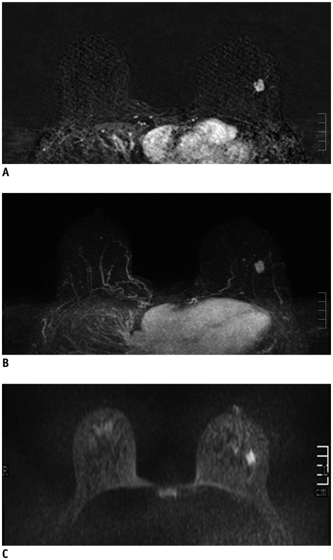
Fig. 1 40-year-old woman with dense breast tissue.FAST (A) and MIP (B) of MRI show 11-mm homogeneously enhancing mass with circumscribed margins in left breast, which was classified as probably benign (BI-RADS 3). However, DWI (C) shows high signal (low signal on apparent diffusion coefficient), which was classified as malignant (BI-RADS 4), and biopsy yielded invasive ductal carcinoma.BI-RADS = Breast Imaging-Reporting and Data System, DWI = diffusion-weighted imaging, FAST = first post-contrast subtracted, MIP = maximum-intensity projection
Cited by 4 articles
-
Abbreviated Magnetic Resonance Imaging for Breast Cancer Screening: Concept, Early Results, and Considerations
Eun Sook Ko, Elizabeth A. Morris
Korean J Radiol. 2019;20(4):533-541. doi: 10.3348/kjr.2018.0722.Age of Data in Contemporary Research Articles Published in Representative General Radiology Journals
Ji Hun Kang, Dong Hwan Kim, Seong Ho Park, Jung Hwan Baek
Korean J Radiol. 2018;19(6):1172-1178. doi: 10.3348/kjr.2018.19.6.1172.Identification of Preoperative Magnetic Resonance Imaging Features Associated with Positive Resection Margins in Breast Cancer: A Retrospective Study
Jung-Hyun Kang, Ji Hyun Youk, Jeong-Ah Kim, Hye Mi Gweon, Na Lae Eun, Kyung Hee Ko, Eun Ju Son
Korean J Radiol. 2018;19(5):897-904. doi: 10.3348/kjr.2018.19.5.897.Kinetic Features of Invasive Breast Cancers on Computer-Aided Diagnosis Using 3T MRI Data: Correlation with Clinical and Pathologic Prognostic Factors
Sung Eun Song, Kyu Ran Cho, Bo Kyoung Seo, Ok Hee Woo, Seung Pil Jung, Deuk Jae Sung
Korean J Radiol. 2019;20(3):411-421. doi: 10.3348/kjr.2018.0587.
Reference
-
1. Sprague BL, Gangnon RE, Burt V, Trentham-Dietz A, Hampton JM, Wellman RD, et al. Prevalence of mammographically dense breasts in the United States. J Natl Cancer Inst. 2014; 106:dju255. PMID: 25217577.
Article2. Ko SY, Kim EK, Kim MJ, Moon HJ. Mammographic density estimation with automated volumetric breast density measurement. Korean J Radiol. 2014; 15:313–321. PMID: 24843235.
Article3. Lee EH, Kim KW, Kim YJ, Shin DR, Park YM, Lim HS, et al. Performance of screening mammography: a report of the alliance for breast cancer screening in Korea. Korean J Radiol. 2016; 17:489–496. PMID: 27390540.
Article4. Graf O, Berg WA, Sickles EA. Large rodlike calcifications at mammography: analysis of morphologic features. AJR Am J Roentgenol. 2013; 200:299–303. PMID: 23345349.
Article5. Pollán M, Ascunce N, Ederra M, Murillo A, Erdozáin N, Alés-Martínez J, et al. Mammographic density and risk of breast cancer according to tumor characteristics and mode of detection: a Spanish population-based case-control study. Breast Cancer Res. 2013; 15:R9. PMID: 23360535.
Article6. Boyd NF, Guo H, Martin LJ, Sun L, Stone J, Fishell E, et al. Mammographic density and the risk and detection of breast cancer. N Engl J Med. 2007; 356:227–236. PMID: 17229950.
Article7. Mandelson MT, Oestreicher N, Porter PL, White D, Finder CA, Taplin SH, et al. Breast density as a predictor of mammographic detection: comparison of interval- and screen-detected cancers. J Natl Cancer Inst. 2000; 92:1081–1087. PMID: 10880551.
Article8. Hooley RJ, Greenberg KL, Stackhouse RM, Geisel JL, Butler RS, Philpotts LE. Screening US in patients with mammographically dense breasts: initial experience with Connecticut Public Act 09-41. Radiology. 2012; 265:59–69. PMID: 22723501.
Article9. Seo M, Cho N, Bae MS, Koo HR, Kim WH, Lee SH, et al. Features of undiagnosed breast cancers at screening breast MR imaging and otential utility of computer-aided evaluation. Korean J Radiol. 2016; 17:59–68. PMID: 26798217.10. Berg WA. How well does supplemental screening magnetic resonance imaging work in high-risk women? J Clin Oncol. 2014; 32:2193–2196. PMID: 24934782.
Article11. Moschetta M, Telegrafo M, Rella L, Capolongo A, Stabile Ianora AA, Angelelli G, et al. MR evaluation of breast lesions obtained by diffusion-weighted imaging with background body signal suppression (DWIBS) and correlations with histological findings. Magn Reson Imaging. 2014; 32:605–609. PMID: 24721005.
Article12. Kul S, Cansu A, Alhan E, Dinc H, Gunes G, Reis A. Contribution of diffusion-weighted imaging to dynamic contrast-enhanced MRI in the characterization of breast tumors. AJR Am J Roentgenol. 2011; 196:210–217. PMID: 21178069.
Article13. Harvey SC, Di Carlo PA, Lee B, Obadina E, Sippo D, Mullen L. An abbreviated protocol for high-risk screening breast MRI saves time and resources. J Am Coll Radiol. 2016; 13:374–380. PMID: 26521970.
Article14. Moschetta M, Telegrafo M, Rella L, Stabile Ianora AA, Angelelli G. Abbreviated combined MR protocol: a new faster strategy for characterizing breast lesions. Clin Breast Cancer. 2016; 16:207–211. PMID: 27108218.
Article15. Grimm LJ, Soo MS, Yoon S, Kim C, Ghate SV, Johnson KS, et al. Abbreviated screening protocol for breast MRI: a feasibility study. Acad Radiol. 2015; 22:1157–1162. PMID: 26152500.16. Morris EA. Rethinking breast cancer screening: ultra FAST breast magnetic resonance imaging. J Clin Oncol. 2014; 32:2281–2283. PMID: 24958827.
Article17. Roubidoux MA, Bailey JE, Wray LA, Helvie MA. Invasive cancers detected after breast cancer screening yielded a negative result: relationship of mammographic density to tumor prognostic factors. Radiology. 2004; 230:42–48. PMID: 14695385.
Article18. Tudorica LA, Oh KY, Roy N, Kettler MD, Chen Y, Hemmingson SL, et al. A feasible high spatiotemporal resolution breast DCE-MRI protocol for clinical settings. Magn Reson Imaging. 2012; 30:1257–1267. PMID: 22770687.
Article19. Berg WA, Zhang Z, Lehrer D, Jong RA, Pisano ED, Barr RG, et al. Detection of breast cancer with addition of annual screening ultrasound or a single screening MRI to mammography in women with elevated breast cancer risk. JAMA. 2012; 307:1394–1404. PMID: 22474203.
Article20. Heacock L, Melsaether AN, Heller SL, Gao Y, Pysarenko KM, Babb JS, et al. Evaluation of a known breast cancer using an abbreviated breast MRI protocol: correlation of imaging characteristics and pathology with lesion detection and conspicuity. Eur J Radiol. 2016; 85:815–823. PMID: 26971429.
Article21. Mango VL, Morris EA, David Dershaw D, Abramson A, Fry C, Moskowitz CS, et al. Abbreviated protocol for breast MRI: are multiple sequences needed for cancer detection? Eur J Radiol. 2015; 84:65–70. PMID: 25454099.
Article22. Heywang SH, Wolf A, Pruss E, Hilbertz T, Eiermann W, Permanetter W. MR imaging of the breast with Gd-DTPA: use and limitations. Radiology. 1989; 171:95–103. PMID: 2648479.
Article23. Kuhl CK, Schrading S, Strobel K, Schild HH, Hilgers RD, Bieling HB. Abbreviated breast magnetic resonance imaging (MRI): first postcontrast subtracted images and maximum-intensity projection-a novel approach to breast cancer screening with MRI. J Clin Oncol. 2014; 32:2304–2310. PMID: 24958821.
Article24. Yoon H, Yoon D, Yun M, Choi JS, Park VY, Kim EK, et al. Metabolomics of breast cancer using high-resolution magic angle spinning magnetic resonance spectroscopy: correlations with 18F-FDG positron emission tomography-computed tomography, dynamic contrast-enhanced and diffusion-weighted imaging MRI. PLoS One. 2016; 11:e0159949. PMID: 27459480.
Article
- Full Text Links
-
- Actions
-
Cited
- CITED
-
- Close
- Share
-
- Similar articles
-
- Abbreviated Breast Magnetic Resonance Imaging: Background, Evidence From Studies, and Future Considerations
- Use of Abbreviated Magnetic Resonance Imaging in Breast Cancer Screening
- Abbreviated Magnetic Resonance Imaging for Breast Cancer Screening: Concept, Early Results, and Considerations
- Distribution of dense breasts using screening mammography in Korean women: a retrospective observational study
- Automated Breast Ultrasound Screening for Dense Breasts
