A Review of the Use of Cardiac Computed Tomography for Evaluating the Mitral Valve before and after Mitral Valve Repair
- Affiliations
-
- 1Department of Thoracic and Cardiovascular Surgery, Chonbuk National University Medical School, Research Institute of Clinical Medicine of Chonbuk National University, Biomedical Research Institute of Chonbuk National University Hospital, Jeonju 54907, Korea.
- 2Department of Radiology, Research Institute of Clinical Medicine of Chonbuk National University, Biomedical Research Institute of Chonbuk National University Hospital, Institute for Medical Sciences of Chonbuk National University Medical School, Jeonju 54907, Korea. key8078@naver.com
- 3Department of Radiology, Kangbuk Samsung Hospital, Sungkyunkwan University School of Medicine, Seoul 03181, Korea.
- KMID: 2427214
- DOI: http://doi.org/10.3348/kjr.2017.18.5.773
Abstract
- The role of cardiac computed tomography (CT) for evaluating the mitral valve (MV) has been limited since echocardiography is the main method of evaluation. However, recent advances in cardiac CT have enable detailed evaluation of the anatomy and geometry of the MV. We describe assessments of the anatomy and coaptation geometric parameters of normal MVs, and also review repair of diseased/damaged MV. We also discuss pre- and post-surgical imaging of MV pathology using cardiac CT and various CT images. We found that cardiac CT could be used as an alternative imaging modality to echocardiography for pre-operative MV evaluation and to predict clinical outcomes following repair.
Keyword
MeSH Terms
Figure
-

Fig. 1 Normal CT images of MV.A. Reconstructed short-axis view (en face view) showing each MV leaflet scallop (A1–A3 and P1–P3). In lateral portion of the mitral annulus, LAA and LCx are shown, and in anterior portion, aorta is seen. These landmarks are used for performing reconstruction in en face view of MV. B. Reconstructed three-chamber view (sagittal view) showing anterior leaflet (arrow) and posterior leaflet (dotted arrow) of MV. Fibrous continuity (arrowhead) is identified between aorta and anterior mitral leaflet. C. Reconstructed two-chamber view (coronal view) showing leaflets (arrow), chordae tendineae (dotted arrow), and papillary muscles (asterisks). Posterior leaflet of MV is divided into P1 (lateral), P2 (middle), and P3 (medial) scallop, respectively, opposing segments of anterior leaflet are named A1 (lateral), A2 (middle), and A3 (medial) scallops, respectively. AL = anterolateral commissure, Ao = aorta, CT = computed tomography, LA = left atrium, LAA = left atrial appendage, LCx = left circumflex artery, LV = left ventricle, MV = mitral valve, PM = posteromedial commissure
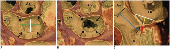
Fig. 2 MV coaptation geometry.A, B. 3D volume-rendered CT images (en face view) showing mitral annulus (arrows). Intercommissural distance (green line) and septo-lateral distance (white line) can be measured in systolic (A) and diastolic phases (B). C. Geometric parameters such as septo-lateral distance (white line), tenting height (yellow line), coaptation length (red line), and lateral displacement of coaptation (blue line) can be observed and measured in three-chamber view of MV by using 3D volume-rendered CT image. 3D = three-dimensional
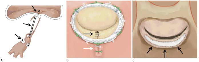
Fig. 3 Illustration of MV repair.A. Artificial neochordae with polytetrafluoroethylene sutures (black arrows). B. Carpentier-Edwards Physio ring with quadrangular resection of posterior mitral leaflet (black arrow) and plication sutures of posterior mitral annulus (white arrow). C. Pericardial patch leaflet augmentation (black arrows).
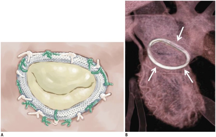
Fig. 4 Carpentier-Edwards Physio annuloplasty ring.Illustrative image (A) and CT image (B) showing high-density ring structure that surrounds annulus, i.e., Carpentier-Edwards Physio annuloplasty ring (white arrows).

Fig. 5 Cosgrove-Edwards annuloplasty system.Illustrative image (A) and CT image (B) showing Cosgrove-Edwards annuloplasty system (black arrows) that partially surrounds mitral annulus.
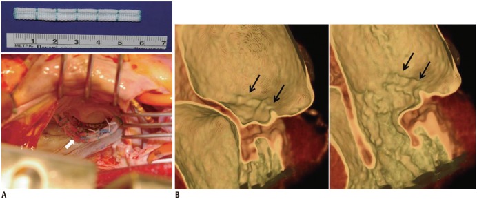
Fig. 6 Intraoperative photograph and post-operative CT images after posterior mitral annuloplasty with ML strip.A. Intraoperative photograph showing posterior mitral annuloplasty strip (white arrow) placed on atrial wall 5.0 mm above posterior annulus, which decreased posterior annular circumference and spared anterior annulus and both commissures. B. Ridge-like lesion (black arrows) was identified on post-operative CT images during systolic and diastolic phases. ML strip = Mitra-Lift® strip
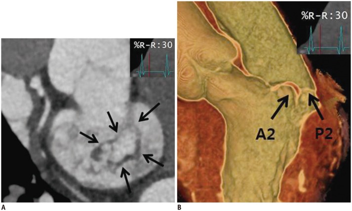
Fig. 7 52-year-old man who presented with chest discomfort.Pre-operative CT images of MV. MV (arrows) was reconstructed by using multiplanar reformat display technique and 3D volume-rendered image with thin-slap reformation technique. En face view (A) and three-chamber view of 3D volume-rendered image (B) at level of MV during systolic phase showing prolapse at A2–A3 level of anterior mitral leaflet. Color Doppler echocardiography showed moderate degree of eccentric mitral regurgitation.
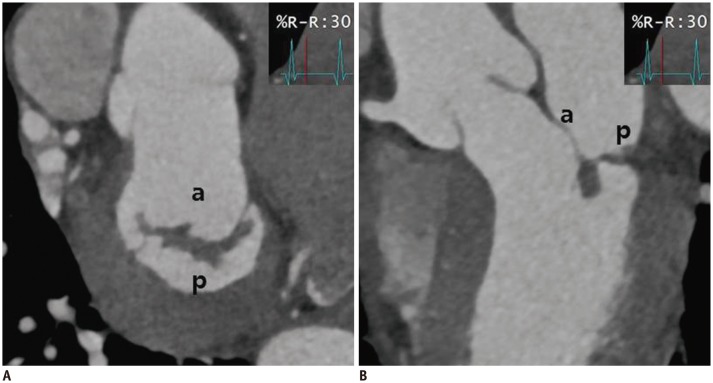
Fig. 8 Cardiac CT images after MV repair due to mitral regurgitation caused by MV prolapse.En face view (A) and three-chamber view (B) at level of mitral valve during systolic phase showing well functioning mitral valve without coaptation defect. a = anterior leaflet of MV, p = posterior leaflet of MV
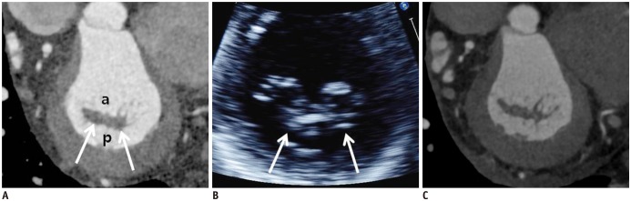
Fig. 9 Pre-operative and post-operative cardiac CT and echocardiographic images of 59-year-old man who presented with palpitation.A. En face views at level of MV (white arrows) during systolic phase showing diffuse valvular thickening without definite mitral stenosis or regurgitation. Coaptation failure was not likely according to multiplanar reformatted images. B. Diffuse valvular thickening (white arrows) was also shown on echocardiography. Color Doppler echocardiography showed moderate degree of mitral regurgitation. C. Diffuse valvular thickening decreased after MV repair, but post-operative cardiac CT still showed small coaptation defect in MV.
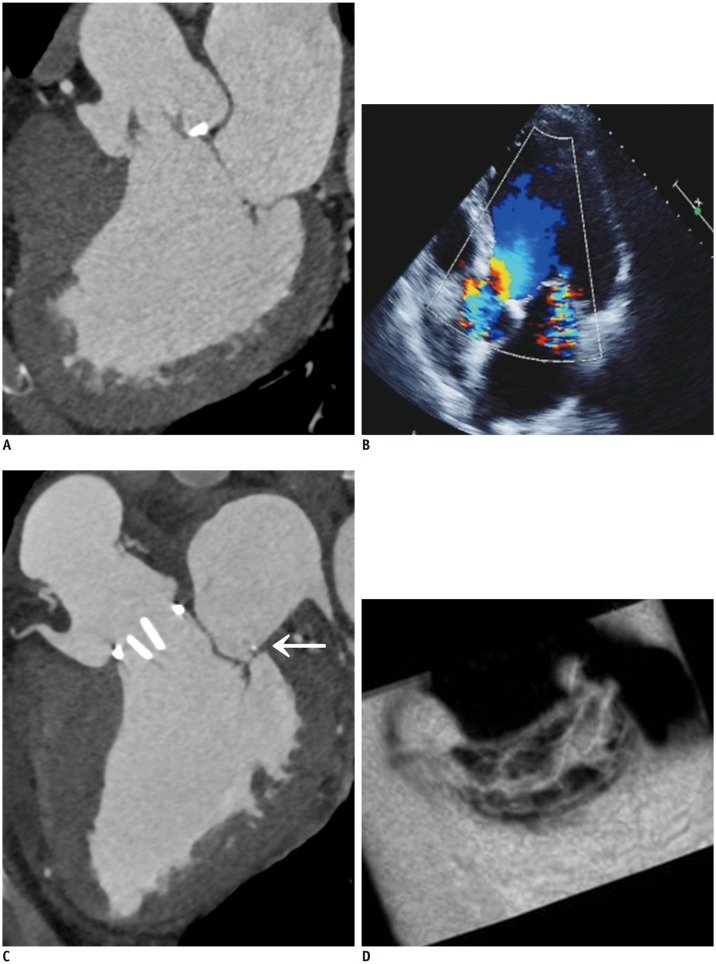
Fig. 10 63-year-old man who presented with exertional dyspnea.A. Pre-operative cardiac CT of multiplanar reformatted images in three-chamber view of MV. B. Echocardiographic image is showing functional mitral regurgitation combined with bicuspid aortic valve with eccentric AR and aneurysmal dilatation of ascending aorta. C, D. Aortic valve replacement and ascending aortic aneurysm resection with graft interposition and Cosgrove-Edwards annuloplasty (C, arrow) were performed for functional mitral regurgitation. Post-operative cardiac CT of multiplanar reformatted images in three-chamber view (C) and en face view of MV (D). En face view of MV show well-functioning MV with good coaptation (D). AR = atrial regurgitation
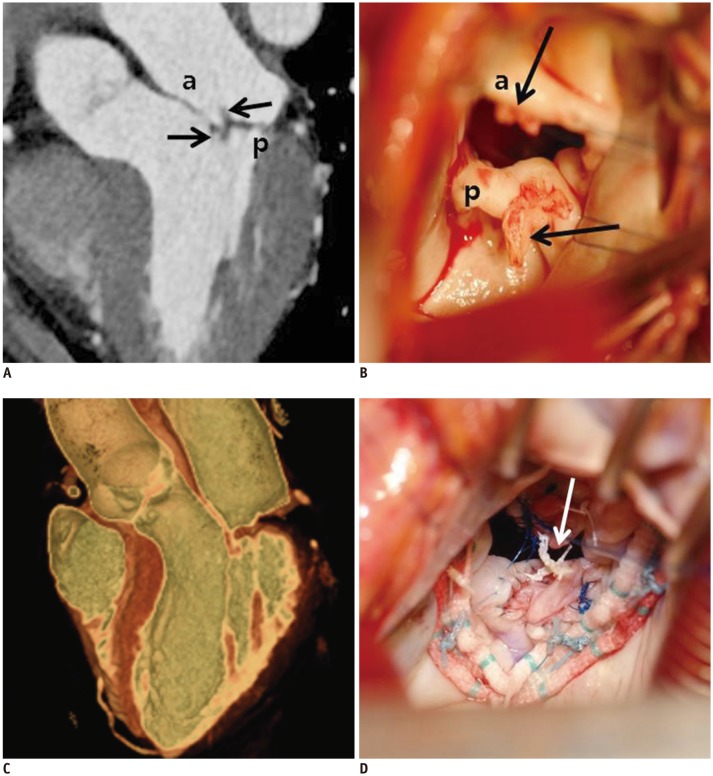
Fig. 11 57-year-old woman who presented with necrosis of left toe and finger.Pre-operative cardiac CT was obtained to evaluate MV and rule out infective endocarditis with vegetation.A. Three-chamber view at level of MV was reconstructed. There were two small soft tissue density masses (black arrows) in anterior and posterior leaflets of MV with mild coaptation defect. B. Intraoperative photograph demonstrating two small masses (black arrows) in anterior and posterior leaflets of MV with endocarditis. Pre-operative echocardiography showed thickened MV with shaggy echogenic material on anterior and posterior mitral leaflets and mild mitral regurgitation. C. Cardiac CT after MV repair. MV was reconstructed by using 3D volume-rendered image with thin-slap reformation technique; well-functioning MV without coaptation defect was observed. D. Two small masses were removed with entire layers of leaflet. Leaflet defects were repaired with autologous pericardial patches and posterior mitral annuloplasty. Artificial neochordae (arrow) were replaced. Histological diagnosis of two small masses was confirmed as papillary fibroelastoma.
Cited by 1 articles
-
Utility of Cardiac CT for Preoperative Evaluation of Mitral Regurgitation: Morphological Evaluation of Mitral Valve and Prediction of Valve Replacement
Young Joo Suh, Sak Lee, Byung-Chul Chang, Chi Young Shim, Geu-Ru Hong, Byoung Wook Choi, Young Jin Kim
Korean J Radiol. 2019;20(3):352-363. doi: 10.3348/kjr.2018.0350.
Reference
-
1. Chen JJ, Manning MA, Frazier AA, Jeudy J, White CS. CT angiography of the cardiac valves: normal, diseased, and postoperative appearances. Radiographics. 2009; 29:1393–1412. PMID: 19755602.
Article2. Shanks M, Delgado V, Ng AC, van der Kley F, Schuijf JD, Boersma E, et al. Mitral valve morphology assessment: three-dimensional transesophageal echocardiography versus computed tomography. Ann Thorac Surg. 2010; 90:1922–1929. PMID: 21095337.
Article3. Delgado V, Tops LF, Schuijf JD, de Roos A, Brugada J, Schalij MJ, et al. Assessment of mitral valve anatomy and geometry with multislice computed tomography. JACC Cardiovasc Imaging. 2009; 2:556–565. PMID: 19442940.
Article4. Kim YJ, Yong HS, Kim SM, Kim JA, Yang DH, Hong YJ, et al. Korean guidelines for the appropriate use of cardiac CT. Korean J Radiol. 2015; 16:251–285. PMID: 25741189.
Article5. Dal-Bianco JP, Levine RA. Anatomy of the mitral valve apparatus: role of 2D and 3D echocardiography. Cardiol Clin. 2013; 31:151–164. PMID: 23743068.6. Levine RA, Handschumacher MD, Sanfilippo AJ, Hagege AA, Harrigan P, Marshall JE, et al. Three-dimensional echocardiographic reconstruction of the mitral valve, with implications for the diagnosis of mitral valve prolapse. Circulation. 1989; 80:589–598. PMID: 2766511.
Article7. McCarthy KP, Ring L, Rana BS. Anatomy of the mitral valve: understanding the mitral valve complex in mitral regurgitation. Eur J Echocardiogr. 2010; 11:i3–i9. PMID: 21078837.
Article8. Kaplan SR, Bashein G, Sheehan FH, Legget ME, Munt B, Li XN, et al. Three-dimensional echocardiographic assessment of annular shape changes in the normal and regurgitant mitral valve. Am Heart J. 2000; 139:378–387. PMID: 10689248.
Article9. Watanabe N, Ogasawara Y, Yamaura Y, Wada N, Kawamoto T, Toyota E, et al. Mitral annulus flattens in ischemic mitral regurgitation: geometric differences between inferior and anterior myocardial infarction: a real-time 3-dimensional echocardiographic study. Circulation. 2005; 112(9 Suppl):I458–I462. PMID: 16159863.
Article10. Fedak PW, McCarthy PM, Bonow RO. Evolving concepts and technologies in mitral valve repair. Circulation. 2008; 117:963–974. PMID: 18285577.
Article11. Ormiston JA, Shah PM, Tei C, Wong M. Size and motion of the mitral valve annulus in man. I. A two-dimensional echocardiographic method and findings in normal subjects. Circulation. 1981; 64:113–120. PMID: 7237707.
Article12. Puff A. Ischemic mitral incompetence. New York: Springer;1991.13. Angelini A, Ho SY, Thiene G, Anderson RH. Anatomy of the mitral valve. In : Boudoulas H, Wooley CF, editors. Mitral valve: floppy mitral valve, mitral valve prolapse, mitral valvular regurgitation. 2nd ed. New York: Futura Publishing Company;2000. p. 5–29.14. Carpentier AF, Lessana A, Relland JY, Belli E, Mihaileanu S, Berrebi AJ, et al. The “physio-ring”: an advanced concept in mitral valve annuloplasty. Ann Thorac Surg. 1995; 60:1177–1185. discussion 1185-1186. PMID: 8526596.
Article15. Lam JH, Ranganathan N, Wigle ED, Silver MD. Morphology of the human mitral valve. I. Chordae tendineae: a new classification. Circulation. 1970; 41:449–458. PMID: 5415982.16. Degandt AA, Weber PA, Saber HA, Duran CM. Mitral valve basal chordae: comparative anatomy and terminology. Ann Thorac Surg. 2007; 84:1250–1255. PMID: 17888977.
Article17. Rusted IE, Scheifley CH, Edwards JE. Studies of the mitral valve. I. Anatomic features of the normal mitral valve and associated structures. Circulation. 1952; 6:825–831. PMID: 12998105.
Article18. Mahmood F, Subramaniam B, Gorman JH 3rd, Levine RM, Gorman RC, Maslow A, et al. Three-dimensional echocardiographic assessment of changes in mitral valve geometry after valve repair. Ann Thorac Surg. 2009; 88:1838–1844. PMID: 19932245.
Article19. Muresian H. The clinical anatomy of the mitral valve. Clin Anat. 2009; 22:85–98. PMID: 18773480.
Article20. Noack T, Kiefer P, Ionasec R, Voigt I, Mansi T, Vollroth M, et al. New concepts for mitral valve imaging. Ann Cardiothorac Surg. 2013; 2:787–795. PMID: 24349983.21. Jensen H, Jensen MO, Smerup MH, Vind-Kezunovic S, Ringgaard S, Andersen NT, et al. Impact of papillary muscle relocation as adjunct procedure to mitral ring annuloplasty in functional ischemic mitral regurgitation. Circulation. 2009; 120(11 Suppl):S92–S98. PMID: 19752392.
Article22. Carpentier A. Cardiac valve surgery--the “French correction”. J Thorac Cardiovasc Surg. 1983; 86:323–337. PMID: 6887954.
Article23. Nishimura RA, Otto CM, Bonow RO, Carabello BA, Erwin JP 3rd, Guyton RA, et al. 2014 AHA/ACC guideline for the management of patients with valvular heart disease: a report of the American College of Cardiology/American Heart Association Task Force on Practice Guidelines. J Thorac Cardiovasc Surg. 2014; 148:e1–e132. PMID: 24939033.24. Zhou YX, Leobon B, Berthoumieu P, Roux D, Glock Y, Mei YQ, et al. Long-term outcomes following repair or replacement in degenerative mitral valve disease. Thorac Cardiovasc Surg. 2010; 58:415–421. PMID: 20922625.
Article25. Goldstone AB, Woo YJ. Surgical treatment of the mitral valve. In : Sellke FW, del Nido PJ, Swanson SJ, editors. Sabiston and Spencer surgery of the chest. 9th ed. Philadelphia: Saunders Elsevier;2016. p. 1384–1429.26. von Oppell UO, Mohr FW. Chordal replacement for both minimally invasive and conventional mitral valve surgery using premeasured Gore-Tex loops. Ann Thorac Surg. 2000; 70:2166–2168. PMID: 11156150.
Article27. David TE, Omran A, Armstrong S, Sun Z, Ivanov J. Long-term results of mitral valve repair for myxomatous disease with and without chordal replacement with expanded polytetrafluoroethylene sutures. J Thorac Cardiovasc Surg. 1998; 115:1279–1285. PMID: 9628669.
Article28. Braunberger E, Deloche A, Berrebi A, Abdallah F, Celestin JA, Meimoun P, et al. Very long-term results (more than 20 years) of valve repair with Carpentier’s techniques in nonrheumatic mitral valve insufficiency. Circulation. 2001; 104(12 Suppl 1):I8–I11. PMID: 11568021.
Article29. Zegdi R, Khabbaz Z, Chauvaud S, Latremouille C, Fabiani JN, Deloche A. Posterior leaflet extension with an autologous pericardial patch in rheumatic mitral insufficiency. Ann Thorac Surg. 2007; 84:1043–1044. PMID: 17720438.
Article30. van Herwerden LA, Taams MA, Bos E. Repair of commissural prolapse by extended leaflet sliding. Ann Thorac Surg. 1994; 57:387–390. PMID: 8311601.
Article31. Cosgrove DM 3rd, Arcidi JM, Rodriguez L, Stewart WJ, Powell K, Thomas JD. Initial experience with the Cosgrove-Edwards Annuloplasty System. Ann Thorac Surg. 1995; 60:499–503. discussion 503-504. PMID: 7677471.
Article32. Kim JH, Kim KH, Choi JB, Kuh JH. Posterior mitral annuloplasty for enhancing mitral leaflet coaptation: using a strip designed for placement in the posterior annulus. J Cardiothorac Surg. 2015; 10:164. PMID: 26563309.
Article33. Song MG, Shin JK, Chee HK, Kim JS, Yang HS, Choi JB. Lifting posterior mitral annuloplasty for enhancing leaflet coaptation in mitral valve repair: midterm outcomes. Ann Cardiothorac Surg. 2015; 4:249–256. PMID: 26309826.34. Koo HJ, Yang DH, Oh SY, Kang JW, Kim DH, Song JK, et al. Demonstration of mitral valve prolapse with CT for planning of mitral valve repair. Radiographics. 2014; 34:1537–1552. PMID: 25310416.
Article35. Salis S, Mazzanti VV, Merli G, Salvi L, Tedesco CC, Veglia F, et al. Cardiopulmonary bypass duration is an independent predictor of morbidity and mortality after cardiac surgery. J Cardiothorac Vasc Anesth. 2008; 22:814–822. PMID: 18948034.
Article36. Feuchtner GM, Alkadhi H, Karlo C, Sarwar A, Meier A, Dichtl W, et al. Cardiac CT angiography for the diagnosis of mitral valve prolapse: comparison with echocardiography. Radiology. 2010; 254:374–383. PMID: 20093510.
Article37. Shah RG, Novaro GM, Blandon RJ, Wilkinson L, Asher CR, Kirsch J. Mitral valve prolapse: evaluation with ECG-gated cardiac CT angiography. AJR Am J Roentgenol. 2010; 194:579–584. PMID: 20173131.
Article38. Beaudoin J, Thai WE, Wai B, Handschumacher MD, Levine RA, Truong QA. Assessment of mitral valve adaptation with gated cardiac computed tomography: validation with three-dimensional echocardiography and mechanistic insight to functional mitral regurgitation. Circ Cardiovasc Imaging. 2013; 6:784–789. PMID: 23873402.39. Morris MF, Maleszewski JJ, Suri RM, Burkhart HM, Foley TA, Bonnichsen CR, et al. CT and MR imaging of the mitral valve: radiologic-pathologic correlation. Radiographics. 2010; 30:1603–1620. PMID: 21071378.
Article40. Chheda SV, Srichai MB, Donnino R, Kim DC, Lim RP, Jacobs JE. Evaluation of the mitral and aortic valves with cardiac CT angiography. J Thorac Imaging. 2010; 25:76–85. PMID: 20160607.
Article41. Pham N, Zaitoun H, Mohammed TL, DeLaPena-Almaguer E, Martinez F, Novaro GM, et al. Complications of aortic valve surgery: manifestations at CT and MR imaging. Radiographics. 2012; 32:1873–1892. PMID: 23150846.
Article42. Guo YK, Yang ZG, Ning G, Rao L, Dong L, Pen Y, et al. Isolated mitral regurgitation: quantitative assessment with 64-section multidetector CT--comparison with MR imaging and echocardiography. Radiology. 2009; 252:369–376. PMID: 19451543.
Article43. Suh YJ, Kim YJ, Hong YJ, Lee HJ, Hur J, Im DJ, et al. Measurement of opening and closing angles of aortic valve prostheses in vivo using dual-source computed tomography: comparison with those of manufacturers’ in 10 different types. Korean J Radiol. 2015; 16:1012–1023. PMID: 26356549.44. Halpern EJ. Clinical applications of cardiac CT angiography. Insights Imaging. 2010; 1:205–222. PMID: 22347917.
Article45. Salgo IS, Gorman JH 3rd, Gorman RC, Jackson BM, Bowen FW, Plappert T, et al. Effect of annular shape on leaflet curvature in reducing mitral leaflet stress. Circulation. 2002; 106:711–717. PMID: 12163432.
Article
- Full Text Links
-
- Actions
-
Cited
- CITED
-
- Close
- Share
-
- Similar articles
-
- Mitral Valve Repair
- Congenital Double-Orifice Mitral Valve with Mitral Valve Prolapse and Severe Mitral Regurgitation
- Unique Congenital Malformation of the Mitral Valve Associated with Anomalous Coronary Arteries and Stroke
- Massive Hemoptysis due to Acute Mitral Regurgitation with Sporadic Primary Mitral Valve Prolapse
- Mitral Valve Repair for Barlow’s Disease with Mitral Annular and Subvalvular Calcification: A Case Report
