Analysis of the Effective Fraction of Sun Ginseng Extract in Selenite Induced Cataract Rat Model
- Affiliations
-
- 1Department of Ophthalmology, Seoul National University College of Medicine, Seoul, Korea. jjhlee@plaza.snu.ac.kr
- 2Department of Ophthalmology, The Armed Forces Capital Hospital, Seongnam, Korea.
- 3Department of Ophthalmology, Soonchunhyang University College of Medicine, Cheonan, Korea.
- 4Department of Ophthalmology, Incheon Medical Center, Incheon, Korea.
- 5Artificial Eye Center of Clinical Research Institute, Seoul National University Hospital, Seoul, Korea.
- 6Seoul National University College of Pharmacy, Seoul, Korea.
- 7Department of Ophthalmology, Seoul National University Bundang Hospital, Seongnam, Korea.
Abstract
- PURPOSE
To compare the protective effects of saponin and non-saponin Sun-ginseng extract fractions in a selenite-induced rat cataract model.
METHODS
A total of 101 Sprague-Dawley rat pups were divided into four groups by treatment: Sun-ginseng, saponin fraction, non-saponin fraction, and control. For induction of cataracts, sodium selenite 15 nmol/g was injected subcutaneously in 13 day-old rat pups. Sun-ginseng extract 100 microgram/g (Group I, Ginseng Science, Seoul, Korea), saponin fraction 100 microgram/g (Group II), non-saponin fraction 100 microgram/g (Group III), and phosphate buffered saline (Control group) were injected intraperitoneally every two days for a total of seven injections. The rats were sacrified and their lenses were dissected and photographed at day 7 and 14, and the cataracts were graded according to the ratio of the cataract area to the total lens area. The blind method was used for the evaluation of the cataract area.
RESULTS
At day 14, cataract formation rates (CFR) were 33.3% in group I, 76.4% in group II, 41.2% in group III, and 77.7% in the control group. The mean cataract area (MCA) was 13.4+/-20.8% in group I, 14.4+/-11.7% in group II, 5.7+/-7.7% in group III, and 15.8+/-12.1% in the control group. Group III showed statistically significant results compared with those of control group (CFR p=0.001, MCA p=0.001). We observed significantly lower incidence and smaller mean cataract area in Group I and Group III at day 7 compared with the control group (Group I, CFR p=0.018; Group III, CFR p=0.032, MCA p=0.005).
CONCLUSIONS
The protective effects of Sun-ginseng extract are caused by the components in the non-saponin fraction, not by those in the saponin fraction, in a selenite-induced cataract rat model.
Keyword
MeSH Terms
Figure
-

Figure 1. The methods of image analysis for lens area measurement. The cataractous lens area and total lens area was analyzed 2-dimensionally on photos of dissected lens using NIH ImageJ software.
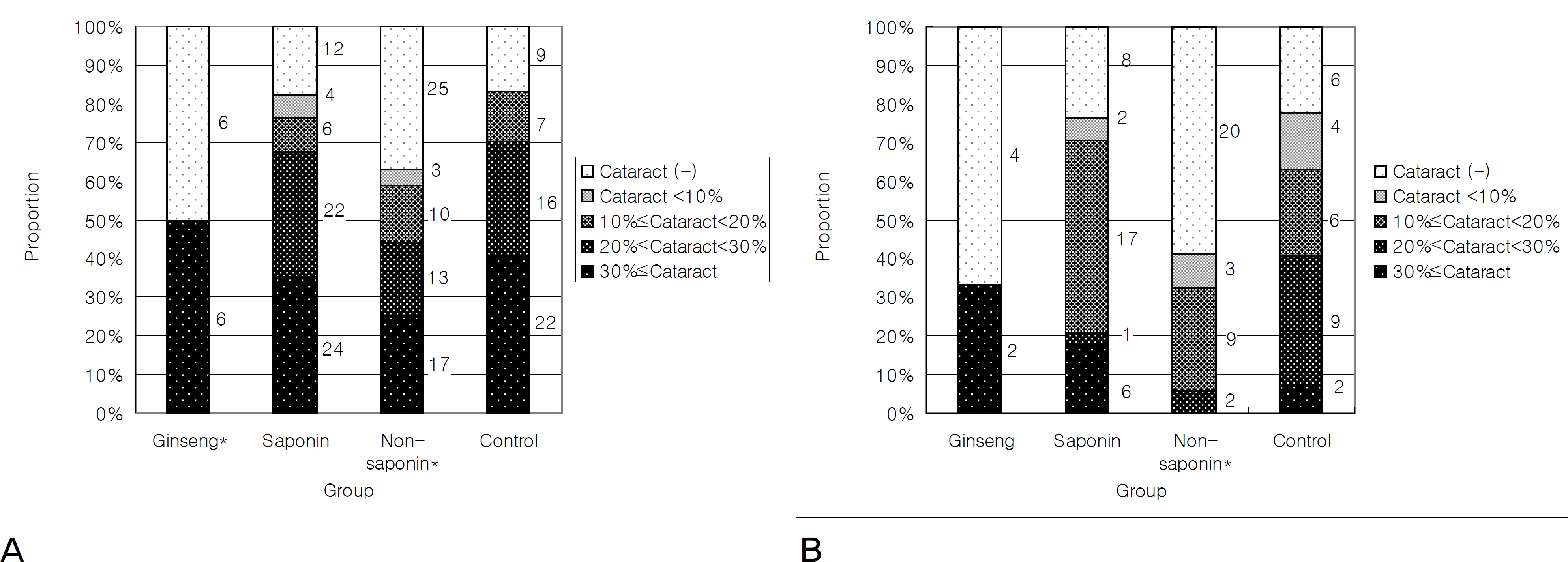
Figure 2. The protective effect of components of ginseng extract in selenite-induced cataract in the rat model. (A) Proportion according to the cataract grade at 1 week after sodium selenite injection. Numbers show total numbers of eyes in each grade. Ginseng group (Group I, p=0.018) and Non-saponin group (Group III, p=0.032) showed lower cataract grade significantly compared with the control group using chi-squre test. (B) Proportion according to the cataract grade at 2 week after sodium selenite injection. Numbers show total numbers of eyes in each grade. Ginseng group (p=0.064) showed borderline difference but Non-saponin group (p=0.001) showed significantly lower cataract grade compared with the control group using chi-squre test. Ginseng: ginseng (sun-sam) extract 100 μg/g every other day injection group; Saponin: saponin component of ginseng extract 100 μg/g every other day injection group; Non-saponin: non-saponin component of ginseng extract 100 μg/g every other day injection group; Control (positive control): PBS every other day injection group. Asterisk (*) indicates statistical significance compared with control group.

Figure 3. The mean area of the cataractous lens to the entire lens. It showed the non-saponin component of ginseng extract group has the protective effect of cataract formation in selenite-induced cataract in the rat model. This graph showed the significantly lower cataract area in non-saponin group (Group III) compared with the control group using Mann-Whitney U test. Mann-Whitney U test was used because the cataract area distribution did not follow the standard curve due to the portions of lenses without cataract. Asterisk (*) indicates statistical significance compared with positive control group.
Reference
-
References
1. Sue Menko A. Lens Epithelial Cell Differentiation. Exp Eye Res. 2002; 75:485–90.
Article2. Bloemendal H, de Jong W, Jaenicke R, et al. Ageing and vision: abdominal, stability and function of lens crystallins. Prog Biophys Mol Biol. 2004; 86:407–85.3. Beebe DC. The lens. Kaufman PL, Alm A, editors. Adler's Physiology of The Eye. 10th ed.St. Louis: Mosby;2002. chap. 5.
Article4. Hejtmancik JF, Piatigorsky J. Lens proteins and their molecular biology. Albert DM, Jakobiec FA, editors. Principles and Practice of Ophthalmology. 2nd ed.Philadelphia: Saunders;2000. 2:chap. 101.
Article5. Palmquist BM, Philipson B, Barr PO. Nuclear cataract and myopia during hyperbaric oxygen therapy. Br J Ophthalmol. 1984; 68:113–7.
Article6. Babizhayev MA, Deyev AI, Lindgerg LF. Lipid peroxidation as a abdominal cause of cataract. Mech Ageing Dev. 1988; 44:69–89.7. Babizhayev MA, Deyev AI. Lens opacity induced by lipid abdominal products as a model of cataract associated with retinal disease. Biochim Biophys Acta. 1989; 1004:124–33.8. Beckman KB, Ames BN. The free radical theory of aging matures. Physiol Rev. 1998; 78:547–81.
Article9. Ostadalova I, Babicky A, Obenberger J. Cataract induced by abdominal of a single dose of sodium selenite to suckling rats. Experientia. 1978; 34:222–3.10. Shearer TR, Ma H, Fukiage C, Azuma M. Selenite nuclear cataract: abdominal of the model. Mol Vis. 1997; 3:8–16.11. David LL, Shearer TR. State of sulfhydryl in selenite cataract. Toxicol Appl Pharmacol. 1984; 74:109–15.
Article12. Wendel A. Selenium in biology and medicine. 1st ed.Vol. 1. New York: Springer-Verlag;1989. p. 70–3.13. Choi KT. Botanical characteristics, pharmacological effects and me-dicinal components of Korean Panax ginseng C A Meyer. Acta Pharmacol Sin. 2008; 29:1109–18.14. Brekhman II, Dardymov IV. New substances of plant origin which abdominal nonspecific resistance. Annu Rev Pharmacol. 1969; 9:419–30.15. Oura H, Oita Y. Effect of Korean red ginseng powder on the survival rate of rat. Ginseng Rev. 1989; 1:228–37.16. Jung MS. Inhibitory Effect of Sun Ginseng Extract on Cataractogenesis in Rat [dissertation]. Cheongju, Korea: Chungbuk National University;2009. p. 1–39.17. Kim WY, Kim JM, Han SB, et al. Steaming of ginseng at high abdominal enhances biological activity. J Nat Prod. 2000; 63:1702–4.18. Kim SI, Lee YH, Kang KS, et al. 10-Acetylpanaxytriol, a new cytotoxic polyacetylene from Panax ginseng. Yakhak Hoeji. 1989; 33:118.19. Ahn BZ, Kim SI, Lee YH. Acetylpanaxydol and panaxydol-chlorohydrin, two new polyenes from Korean ginseng with cytotoxic activity against L1210 cells. Arch Pharm (Weinheim). 1989; 322:223–6.20. Keum YS, Park KK, Lee JM, et al. Antioxidant and Anti-Tumor Promoting Activities of the Methanol Extract of Heat-Processed Ginseng. Cancer Lett. 2000; 150:41–8.
Article21. Lee YJ, Kim HY, Kang KS, et al. The Chemical and Hydroxyl Radical Scavenging Activity Changes of Ginsenoside-Rb1 by Heat Processing. Bioorg Med Chem Lett. 2008; 18:4515–20.
Article22. Kang KS, Kim HY, Baek SH, et al. Study on the hydroxyl radical abdominal activity changes of ginseng and ginsenoside-Rb2 by heat processing. Biol Pharm Bull. 2007; 30:724–8.23. Zhang JT, Qu ZW, Liu Y, Deng HL. Preliminary study on antiamnestic mechanism of ginsenoside Rg1 and Rb1. Clin Med J. 1990; 103:932–8.24. Yun TK, Lee YS, Lee YH, et al. Anticarcinogenic effect of Panax abdominal C.A. Meyer and identification of active compounds. J Korean Med Sci. 2001; 16:S6–18.25. Lee SY, Kim GT, Roh SH, et al. Proteomic analysis of the abdominal effect of 20S-ginsenoside Rg3 in human colon cancer cell lines. Biosci Biotechnol Biochem. 2009; 73:811–6.
- Full Text Links
-
- Actions
-
Cited
- CITED
-
- Close
- Share
-
- Similar articles
-
- Effect of Glutathione With Sea Tangle Extract on Prevention of Selenite-Induced Cataract Formation in Rats
- Effect of Ginseng Extract on Male Rat Sexual Behavior
- An Experimental Study on the Protective Effects of Ginseng Extract to Oxygen Toxicity
- Effect of Ginseng Extract on Blood Lipids and Atherosclerosis
- Effect of Ginseng on the Blood Pressure and Lipid Metabolism, during Development of Hypertension in Spontaneously Hypertensive Rat
