The Effects of Intravitreal Bevacizumab Injection According to the Type of Diabetic Macular Edema
- Affiliations
-
- 1Department of Ophthalmology, Chung-Ang University College of Medicine, Seoul, Korea. hkcho26@cau.ac.kr
Abstract
- PURPOSE
To investigate the effects of intravitreal bevacizumab injection on diabetic macular edema (DME) of different types classified using Optical Coherence Tomography (OCT).
METHODS
A total of 82 eyes with refractory DME were enrolled. The DME was classified into diffuse, cystoid, or serous type based on the OCT findings. All cases had received an intravitreal injection of 1.25 mg bevacizumab each month for three months. Foveal thickness, macular volume, and best corrected visual acuity (BCVA) were measured before and one month after the injection, and the interval changes in these parameters were compared.
RESULTS
The types of DME were classified as follows: diffuse macular edema 50%, cystoid macular edema 31.7%, and serous macular detachment 18.3%. Foveal thickness and total macular volume after intravitreal bevacizumab injection decreased in all types, and the cystoid and serous types showed better response than did the diffuse type with regard to foveal thickness. However, there were no significant differences in the extent of the change in total macular volume or BCVA among the three types of DME.
CONCLUSIONS
There were differences in the therapeutic effects of intravitreal bevacizumab injection among the different types of DME classified using OCT. These differences may be associated with the stabilizing effect of the bevacizumab. This effect was stronger with regard to vascular permeability, the primary factor in the pathogenesis of the cystic and serous types, than with regard to leakage from the microaneurysm, the primary factor in the pathogenesis of the diffuse type. Practical application of bevacizumab to eyes with different DME types will help in further evaluating intravitreal bevacizumab injection as a treatment option for DME.
MeSH Terms
Figure
-
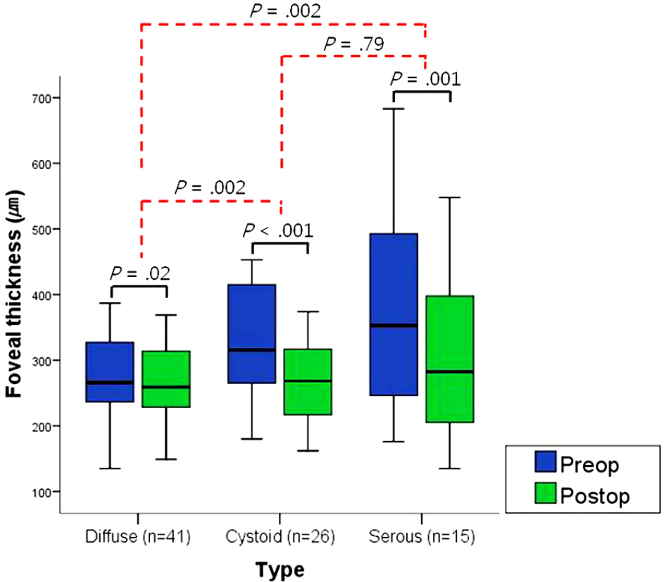
Figure 1. An analysis of the foveal thickness before and 1 month after the third intravitreal bevacizumab injection. Foveal thickness significantly decreased in every type (Wilcoxon's signed rank test, p=.02, p<.001, p=.001, with p<.05 regarded as significant). And the extent of changes in types of cystoid macular edema and serous macular detachment was greater than in diffuse macular edema type (Mann-Whitney U test p=.002, with p<.05 regarded as significant).
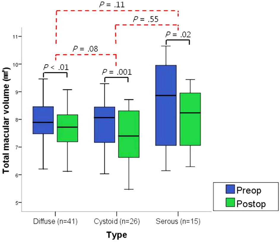
Figure 2. An analysis of total macular volume before and 1 month after the third intravitreal bevacizumab injection. Total macular volume was significantly decreased in every type (Wilcoxon's signed rank test, p=.01, p= .001, p=.02, with p<.05 regarded as significant). But there were no significant differences for the extent of changes among the three types of diabetic macular edema (Mann-Whitney U test p>.05, with p<.05 regarded as significant).
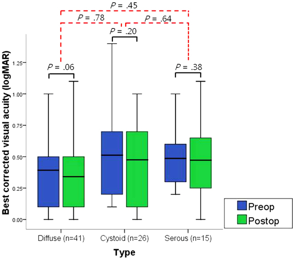
Figure 3. An analysis of the best corrected visual acuity (BCVA) before and 1 month after the third intravitreal bevacizumab injection. There were neither significant differences for changes of BCVA in the three types of macular edema nor for the extent of changes among the three types of diabetic macular edema (Wilcoxon's signed rank test p>.05, Mann-Whitney U test p>.05, with p<.05 regarded as significant).
Reference
-
References
1. Battegay EJ. Angiogenesis: mechanistic insights, neovascular abdominals, and therapeutic prospects. J Mol Med. 1995; 73:333–46.
Article2. De Vries C, Escobedo JA, Ueno H, et al. The fms-like tyrosine kinase, a receptor for vascular endothelial growth factor. Science. 1992; 255:989–91.3. Senger DR, Galli SJ, Dvorak AM, et al. Tumor cells secrete a abdominal permeability factor that promotes accumulation of ascites fluid. Science. 1983; 219:983–5.4. Philipp W, Speicher L, Humpel C. Expression of vascular abdominal growth factor and its receptors in inflamed and vascularized human corneas. Invest Ophthalmol Vis Sci. 2000; 41:2514–22.5. Rosenfeld PJ, Moshfeghi AA, Puliafito CA. Optical coherence abdominal findings after an intravitreal injection of bevacizumab (avastin) for neovascular age-related macular degeneration. Ophthalmic Surg Lasers Imaging. 2005; 36:331–5.6. Haritoglou C, Kook D, Neubauer A, et al. Intravitreal abdominal (avastin) therapy for persistent diffuse diabetic macular edema. Retina. 2006; 26:999–1005.7. Arevalo JF, Fromow-Guerra J, Quiroz-Mercado H, et al. Primary intravitreal bevacizumab (avastin) for diabetic macular edema: abdominal from the Pan-american collaborative retina study group at 6-month follow-up. Ophthalmology. 2007; 114:743–50.8. Klein R, Klein BE, Moss SE, et al. The Wisconsin epidemiologic study of diabetic retinopathy IV: Diabetic macular edema. Ophthalmology. 1984; 91:1464–74.9. Kim HK, Oh TS, Lee SM, Lee JB. The initial fundus examination and severity of diabetic retinopathy at a primary eye clinic. J Korean Ophthalmol Soc. 2005; 46:982–8.10. Early Treatment Diabetic Retinopahty Study Research Group. Photocoagulation for diabetic macular edema. Early treatment abdominal retinopathy study report number 1. Arch Ophthalmol. 1985; 103:1796–806.11. Tano Y, Chandler D, Machemer R. Treatment of intraocular abdominal with intravitreal injection of triamcinolone acetonide. Am J Ophthalmol. 1980; 90:810–6.12. Pendergast SD, Hassan TS, Williams GA, et al. Vitrectomy for diffuse diabetic macular edema associated with a taut premacular posterior hyaloids. Am J Ophthalmol. 2000; 130:178–86.13. Martidis A, Duker JS, Greenberg PB, et al. Intravitreal triamcinolone for refractory diabetic macular edema. Ophthalmology. 2002; 109:920–7.
Article14. Funatsu H, Yamashita H, Ikeda T, et al. Vitreous levels of inter-leukin-6 and vascular endothelial growth factor are related to abdominal macular edema. Ophthalmology. 2003; 110:1690–6.15. Funatsu H, Yamashita H, Ikeda T, et al. Angiotensin II and abdominal endothelial growth factor in the vitreous fluid of patients with diabetic macular edema and other retinal disorders. Am J Ophthalmol. 2002; 133:537–43.16. Chang MW, Kim SW, Oh IK, et al. Intravitreal triamcinolone injection with or without bevacizumab for diabetic macular edema. J Korean Ophthalmol Soc. 2008; 49:1269–74.
Article17. Kim JY, Kweon EY, Lee DW, Cho NC. Results of intravitreal bevacizumab for macular edema with retinal vein occlusion and abdominal macular edema. J Korean Ophthalmol Soc. 2008; 49:1275–82.18. Otani T, Kishi S, Maruyama Y. Patterns of diabetic macular edema with optical coherence tomography. Am J Ophthalmol. 1999; 127:688–93.
Article19. Kang SW, Park CY, Ham DI. The correlation between fluorescein abdominal and optical coherence tomographic features in clinically significant diabetic macular edema. Am J Ophthalmol. 2004; 137:313–22.20. Kim YG, Yu SY, Kwak HW. The effect of intravitreal triamcinolone acetonide injection according to the diabetic macular edema type. J Korean Ophthalmol Soc. 2005; 46:84–9.21. Ferris FL III, Patz A. Macular edema. A complication of diabetic retinopathy. Surv Ophthalmol. 1984; 28:S452–61.22. Antcliff RJ, Marshall J. The pathogenesis of edema in diabetic maculopathy. Semin Ophthalmol. 1999; 14:223–32.
Article23. Cunha-Vaz JG. Diabetic macular edema. Eur J Ophthalmol. 1998; 8:127–30.
Article24. Lobo C, Bernardes R, Faria de Abreu Jr, Cunha-Vaz JG. Novel imaging techniques for diabetic macular edema. Doc Ophthalmol. 1999; 97:341–7.
Article25. Yannoff M, Fine BS, Brucker AJ, Eagle RC Jr. Pathology of abdominal cystoid macular edema. Surv Ophthalmol. 1984; 28:S505–11.26. Fine BS, Brucker AJ. Macular dema. Macular edema and cystoid macular edema. Am J Ophthalmol. 1981; 92:466–81.27. Yoon SC, Lee DY, Nam DH. The effect of intravitreal triamcinolone acetoied injection to the OCT patterns of diabetic macular edema. J Korean Ophthalmol Soc. 2008; 49:1611–8.28. Kim BY, Smith SD, Kaiser PK. Optical coherence tomography patterns of diabetic macular edema. Am J Ophthalmol. 2006; 142:405–12.29. Alkuraya H, Kangave D, Abu El-Asrar AM. The correlation abdominal optical coherence tomographic features and severity of abdominal, macular thickness and visual acuity in diabetic macular edema. Int Ophthalmol. 2005; 26:93–9.30. Cho HY, Lee JH. The correlation between visual acuity and abdominals of diabetic macular edema in OCT images. J Korean Ophthalmol Soc. 2003; 44:2028–34.31. Arevalo JF, Sanchez JG, Wu L, et al. Primary intravitreal abdominal for diffuse diabetic macular edema: the Pan-American Collaborative Retina Study Group at 24 months. Ophthalmology. 2009; 116:1488–97.32. Shimura M, Nakazawa T, Yasuda K, et al. Comparative therapy evaluation of intravitreal bevacizumab and triamcinolone abdominal on persistent diffuse diabetic macular edema. Am J Ophthalmol. 2008; 145:854–61.33. Klein R, Klein BE, Moss SE, et al. The Wisconsin epidemiologic study of diabetic retinopathy IV. Diabetic macular edema. Ophthalmology. 1984; 91:1464–74.34. Lobo CL, Bernardes RC, de Abreu Jr, Cunha-Vaz JG. One-year follow-up of blood-retinal barrier and retinal thickness alteration in patients with type 2 diabetes mellitus and mild nonproliferative retinopathy. Arch Ophthalmol. 2001; 119:1469–74.35. Kojima T, Terasaki H, Nomura H, et al. Vitrectomy for diabetic macular edema: effect of glycemic control(HbA1c), renal function(creatinine) and other local factors. Ophthalmic Res. 2003; 35:192–8.36. Oh IK, Kim HS, Oh J, Huh K. Diurnal variation of macular abdominal in diabetic macular edema. J Korean Ophthalmol Soc. 2005; 46:279–86.37. Chang LK, Koizumi H, Spaide RF. Disruption of the photoreceptor inner segment-outer segment junction in eyes with macular holes. Retina. 2008; 28:969–75.
Article38. Paccola L, Costa RA, Folgosa MS, et al. Intravitreal triamcinolone versus bevacizumab for treatment of refractory diabetic macular edema (IBEME study). Br J Ophthalmol. 2008; 92:76–80.
- Full Text Links
-
- Actions
-
Cited
- CITED
-
- Close
- Share
-
- Similar articles
-
- Macular Hole Formation after Intravitreal Injection of Bevacizumab for Diabetic Macular Edema
- Comparison of Intravitreal Bevacizumab Alone Injection and Intravitreal Combination Low-Dose Bevacizumab-Triamcinolone Injection or Diabetic Macular Edema
- Effects of Intravitreal Bevacizumab Injection in 3 Types of Macular Edema Secondary to Branch Retinal Vein Occlusion
- Comparison of Intravitreal Triamcinolone Versus Bevacizumab in Bilateral Diabetic Macular Edema by Optical Coherence Tomography (OCT) Patterns
- The Effect of Intravitreal Triamcinolone Acetonide Injection according to the Diabetic Macular Edema Type
