Effects of Intravitreal Bevacizumab Injection in 3 Types of Macular Edema Secondary to Branch Retinal Vein Occlusion
- Affiliations
-
- 1Myung-Gok Eye Research Institute, Department of Ophthalmology, Kim's Eye Hospital, College of Medicine, Konyang University, Seoul, Korea. mediceye@kimeye.com
- KMID: 2215953
- DOI: http://doi.org/10.3341/jkos.2012.53.8.1112
Abstract
- PURPOSE
To compare the effects of intravitreal bevacizumab injection (IVB) in 3 types of macular edema secondary to branch retinal vein occlusion (BRVO).
METHODS
Patients with macular edema secondary to BRVO with at least 1-year follow-up after IVB were included in the present retrospective study. The authors classified the macular edema into 3 types according to OCT findings: diffuse macular edema (type 1), cystoid macular edema (type 2), and serous retinal detachment (type 3). The clinical outcome indicators were best corrected visual acuity, central macular thickness (CMT), and total number of injections.
RESULTS
The total injection number was significantly higher in type 2 than type 3 (p = 0.02). Changes in CMT were significantly different between type 2 and type 3 (p = 0.03). CMT decreased significantly after IVB in all types of macular edema.
CONCLUSIONS
Morphologic classification of macular edema with OCT is useful for predicting efficacy of IVB in macular edema secondary to BRVO.
Keyword
MeSH Terms
Figure
-
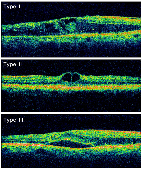
Figure 1 Classification of macular edema in BRVO patients. Type I: diffuse macular edema. Type II: cystoids macular edema. Type III: serous retinal detachment.
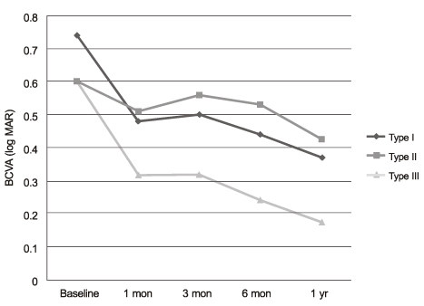
Figure 2 Changes in visual acuity after intravitreal Bevacizumab injection. Type I: diffuse macular edema. Type II: cystoids macular edema. Type III: serous retinal detachment.
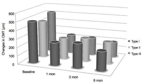
Figure 3 Changes in CMT after intravitreal bevacizumab injection. Type I: diffuse macular edema. Type II: cystoids macular edema. Type III: serous retinal detachment.
Cited by 1 articles
-
Factors Related to Repeatability of Intravitreal Bevacizumab Injections in Branch Retinal Vein Occlusion Macular Edema
Kyung Tae Kang, Yu Cheol Kim, Kwang Soo Kim
J Korean Ophthalmol Soc. 2015;56(10):1580-1585. doi: 10.3341/jkos.2015.56.10.1580.
Reference
-
1. Weinberg D, Dodwell DG, Fern SA. Anatomy of arteriovenous crossings in branch retinal vein occlusion. Am J Ophthalmol. 1990. 109:298–302.2. The Branch Vein Occlusion Study Group. Argon laser photocoagulation for macular edema in branch vein occlusion. Am J Ophthalmol. 1984. 98:271–282.3. Dick SB, Jampol LM, Haller JA. Ryan SJ, editor. Macular edema. Retina. 2001. v. 2:3rd ed. St. Louis: Mosby;chap. 55-7.4. Gutman FA, Zegarra H. Macular edema secondary to occlusion of the retinal veins. Surv Ophthalmol. 1984. 28:suppl. 462–470.5. Finkelstein D. Ryan SJ, editor. Retinal branch vein occlusion. Retina. 1989. v. 2:1st ed. St. Louis: CV Mosby;chap. 18.6. The Branch Vein Occlusion Study Group. Argon laser photocoagulation for macular edema in branch vein occlusion. Am J Ophthalmol. 1984. 98:271–282.7. Jonas JB, Akkoyun I, Kamppeter B, et al. Branch retinal vein occlusion treated by intravitreal triamcinolone acetonide. Eye. 2005. 19:65–71.8. Rabena MD, Pieramici DJ, Castellarin AA, et al. Intravitreal bevacizumab (Avastin) in the treatment of macular edema secondary to branch retinal vein occlusion. Retina. 2007. 27:419–425.9. Tolentino MJ, Miller JW, Gragoudas ES, et al. Intravitreous injections of vascular endothelial growth factor produce retinal ischemia and microangiopathy in an adult primate. Ophthalmology. 1996. 103:1820–1828.10. Noma H, Funatsu H, Yamasaki M, et al. Pathogenesis of macular edema with branch retinal vein occlusion and intraocular levels of vascular endothelial growth factor and interleukin-6. Am J Ophthalmol. 2005. 140:256–261.11. Funk M, Kriechbaum K, Prager F, et al. Intraocular concentrations of growth factors and cytokines in retinal vein occlusion and the effect of therapy with bevacizumab. Invest Ophthalmol Vis Sci. 2009. 50:1025–1032.12. Wu L, Martínez-Castellanos MA, Quiroz-Mercado H, et al. Twelve-month safety of intravitreal injections of bevacizumab (Avastin): results of the Pan-American Collaborative Retina Study Group (PACORES). Graefes Arch Clin Exp Ophthalmol. 2008. 246:81–87.13. Kreutzer TC, Alge CS, Wolf AH, et al. Intravitreal bevacizumab for the treatment of macular oedema secondary to branch retinal vein occlusion. Br J Ophthalmol. 2008. 92:351–355.14. Rabena MD, Pieramici DJ, Castellarin AA, et al. Intravitreal bevacizumab (Avastin) in the treatment of macular edema secondary to branch retinal vein occlusion. Retina. 2007. 27:419–425.15. Matsumoto Y, Freund KB, Peiretti E, et al. Rebound macular edema following bevacizumab (Avastin) therapy for retinal venous occlusive disease. Retina. 2007. 27:426–431.16. Hee MR, Puliafito CA, Wong C, et al. Quantitative assessment of macular edema with optical coherence tomography. Arch Ophthalmol. 1995. 113:1019–1029.17. Otani T, Kishi S, Maruyama Y. Patterns of diabetic macular edema with optical coherence tomography. Am J Ophthalmol. 1999. 127:688–693.18. Noma H, Minamoto A, Funatsu H, et al. Intravitreal levels of vascular endothelial growth factor and interleukin-6 are correlated with macular edema in branch retinal vein occlusion. Graefes Arch Clin Exp Ophthalmol. 2006. 244:309–315.19. Brown GC, Kimmel AS, Magargal LE, et al. Progressive capillary nonperfusion in temporal branch retinal vein obstruction. Ann Ophthalmol. 1989. 21:290–293.20. Gregori NZ, Rattan GH, Rosenfeld PJ, et al. Safety and efficacy of intravitreal bevacizumab (avastin) for the management of branch and hemiretinal vein occlusion. Retina. 2009. 29:913–925.21. Nussenblatt RB, Kaufman SC, Palestine AG, et al. Macular thickening and visual acuity. Measurement in patients with cystoid macular edema. Ophthalmology. 1987. 94:1134–1139.22. Antcliff RJ, Stanford MR, Chauhan DS, et al. Comparison between optical coherence tomography and fundus fluorescein angiography for the detection of cystoid macular edema in patients with uveitis. Ophthalmology. 2000. 107:593–599.23. Jalkh AE, Trempe CL. Macular edema in branch retinal vein occlusion: types and treatment. Ophthalmic Surg. 1989. 20:26–32.24. Rehák J. [Retinal vein occlusion. I. Pathogenesis of circulatory changes]. Cesk Oftalmol. 1993. 49:145–147.25. Finkelstein D. Ischemic macular edema. Recognition and favorable natural history in branch vein occlusion. Arch Ophthalmol. 1992. 110:1427–1434.26. Lee SW, Kim HK, Kim SY. Patterns of macular edema in patients with branch retinal vein occlusion on optical coherence tomography. J Korean Ophthalmol Soc. 2005. 46:969–975.27. Kang SW, Park CY, Ham DI. The correlation between fluorescein angiographic and optical coherence tomographic features in clinically significant diabetic macular edema. Am J Ophthalmol. 2004. 137:313–322.28. Song YB, Park SP. Short-term effects of intravitreal bevacizumab injection and macular edema patterns in branch retinal vein occlusion. J Korean Ophthalmol Soc. 2010. 51:379–385.
- Full Text Links
-
- Actions
-
Cited
- CITED
-
- Close
- Share
-
- Similar articles
-
- The Efficacy of Intravitreal Bevacizumab in the Treatment of Macular Edema
- Intravitreal Bevacizumab Injection for Macular Edema Secondary to Branch Retinal Vein Occlusion: Long-Term Results
- Intravitreal Bevacizumab Injection for Macular Edema Secondary to Branch Retinal Vein Occlusion
- Combined Low Dose Bevacizumab-triamcinolone versus Bevacizumab Single Intravitreal Injection for Branch Retinal Vein Occlusion
- Macular Vessel Density Analysis Using Optical Coherence Tomography Angiography before and after Intravitreal Bevacizumab Injection in Branch Retinal Vein Occlusion
