Undifferentiated embryonal sarcoma of the liver in adult patient: A report of two cases
- Affiliations
-
- 1Surgery Department, Instituto Nacional de Ciencias Médicas y Nutrición Salvador Zubirán, Mexico City, Mexico. ge.sanchez@hotmail.com
- KMID: 2420616
- DOI: http://doi.org/10.14701/ahbps.2018.22.3.269
Abstract
- Primary sarcomas of the liver are unusual neoplasms developing in adults. They constitute a heterogeneous group of neoplasms including undifferentiated embryonal sarcoma. Patients usually present with an abdominal mass and abdominal pain. Case 1: A 53-year-old woman presented with abdominal pain. Computed tomography showed an occupying mass in the right lobule and an intra-auricular multi-lobulated mass suggestive of a secondary deposit. Biopsy of the hepatic lesion revealed undifferentiated embryonal sarcoma. Despite radiotherapy and supportive measures, her overall status progressively worsened until cardiac arrest. Case 2: A 41-year-old woman presented with hepatomegaly. Abdominal imaging showed cystic lesions in the right hepatic lobule with multiple septae. The patient underwent extended right hepatectomy and a histopathological study reported high-grade undifferentiated embryonal sarcoma. Two years after surgery, a new cystic lesion in the surgical site was recorded and chemotherapy was scheduled. The lesion remained stable for three years when disease progression was observed and second-line chemotherapy was initiated. Although undifferentiated embryonal sarcoma of the liver has poor prognosis, early diagnosis is essential to increase the chances of survival. Currently, surgical resection and chemotherapy are the primary treatment modalities.
MeSH Terms
Figure
-
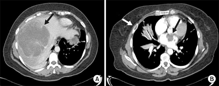
Fig. 1 Computed tomography with intravenous contrast showing a (A) right liver lobule with a 20 cm-sized hypodense lesion (arrow) with irregular borders, a few septa, and centripetal reinforcement without any intra or extrahepatic biliary tract dilation. (B) The heart was augmented in size and the left auricle showed a 39 mm-sized hypodense mass with multi-lobulated borders leading to filling defect (black arrow). In addition, a lytic lesion in the fourth right costal arch was documented (white arrow), both of which suggesting secondary deposits.

Fig. 2 Computed tomography with intravenous contrast that showed a (A) liver enlargement due to the presence of a 26 cm-sized cystic lesion with multiple thick septa (arrow). The lesion showed reinforcement with contrast material without evidence of solid tissue, in addition to compression and displacement of structures in the midline towards the left. Abdominal ultrasound (B) showed a previously described lesion with inner echoes and multiple septa, and compressed intrahepatic vascular structures including portal vein (arrow).

Fig. 3 Computed tomography with intravenous contrast (A) after extended right hepatectomy with post-surgical changes, the left lobule showed homogeneous density and regular form as well as an intrahepatic biliary tract with preserved caliber. Two years later (B) there was a 10 mm-sized hypodense heterogeneous lesion in the anterior portion of the left portal vein without occasional filling defects. The remainder of the liver parenchyma showed no changes. Five years after the initial diagnosis (C) the lesion documented previously showed an increase in size to 30 mm, suggesting disease progression.
Reference
-
1. Liver Cancer Study Group of Japan. Primary liver cancer in Japan. Clinicopathologic features and results of surgical treatment. Ann Surg. 1990; 211:277–287.2. Stocker JT, Ishak KG. Undifferentiated (embryonal) sarcoma of the liver: report of 31 cases. Cancer. 1978; 42:336–348.3. Jiménez Fuertes M, López Andújar R, de Juan Burgueño M, Moya Herráiz A, Sanjuán Rodríguez F, Montalvá Orón E, et al. Sarcoma indiferenciado (embrionario) de hígado del adulto: informe de un caso y revisión de la literatura médica. Gastroenterol Hepatol. 2008; 31:12–17.
Article4. Mori A, Fukase K, Masuda K, Sakata N, Mizuma M, Ohtsuka H, et al. A case of adult undifferentiated embryonal sarcoma of the liver successfully treated with right trisectionectomy: a case report. Surg Case Rep. 2017; 3:19.
Article5. Putra J, Ornvold K. Undifferentiated embryonal sarcoma of the liver: a concise review. Arch Pathol Lab Med. 2015; 139:269–273.
Article6. Sakellaridis T, Panagiotou I, Georgantas T, Micros G, Rontogianni D, Antiochos C. Undifferentiated embryonal sarcoma of the liver mimicking acute appendicitis. Case report and review of the literature. World J Surg Oncol. 2006; 4:9.7. Wei ZG, Tang LF, Chen ZM, Tang HF, Li MJ. Childhood undifferentiated embryonal liver sarcoma: clinical features and immunohistochemistry analysis. J Pediatr Surg. 2008; 43:1912–1919.
Article8. Harman M, Nart D, Acar T, Elmas N. Primary mesenchymal liver tumors: radiological spectrum, differential diagnosis, and pathologic correlation. Abdom Imaging. 2015; 40:1316–1330.
Article9. Pachera S, Nishio H, Takahashi Y, Yokoyama Y, Oda K, Ebata T, et al. Undifferentiated embryonal sarcoma of the liver: case report and literature survey. J Hepatobiliary Pancreat Surg. 2008; 15:536–544.
Article10. Hong WJ, Kang YN, Kang KJ. Undifferentiated embryonal sarcoma in adult liver. Korean J Pathol. 2014; 48:311–314.
Article11. Lee KH, Maratovich MN, Lee KB. Undifferentiated embryonal sarcoma of the liver in an adult patient. Clin Mol Hepatol. 2016; 22:292–295.
Article12. Kiani B, Ferrell LD, Qualman S, Frankel WL. Immunohistochemical analysis of embryonal sarcoma of the liver. Appl Immunohistochem Mol Morphol. 2006; 14:193–197.
Article13. Agaram NP, Baren A, Antonescu CR. Pediatric and adult hepatic embryonal sarcoma: a comparative ultrastructural study with morphologic correlations. Ultrastruct Pathol. 2006; 30:403–408.
Article14. Lauwers GY, Grant LD, Donnelly WH, Meloni AM, Foss RM, Sanberg AA, et al. Hepatic undifferentiated (embryonal) sarcoma arising in a mesenchymal hamartoma. Am J Surg Pathol. 1997; 21:1248–1254.
Article15. Yang L, Chen LB, Xiao J, Han P. Clinical features and spiral computed tomography analysis of undifferentiated embryonic liver sarcoma in adults. J Dig Dis. 2009; 10:305–309.
Article16. Xie ZY, Li LP, Wu WJ, Sun DY, Zhou MH, Zhao YG. Undifferentiated embryonal sarcoma of the liver mistaken for hepatic abscess in an adult. Oncol Lett. 2014; 8:1184–1186.
Article17. Buetow PC, Buck JL, Pantongrag-Brown L, Marshall WH, Ros PR, Levine MS, et al. Undifferentiated (embryonal) sarcoma of the liver: pathologic basis of imaging findings in 28 cases. Radiology. 1997; 203:779–783.
Article18. Kim KA, Kim KW, Park SH, Jang SJ, Park MS, Kim PN, et al. Unusual mesenchymal liver tumors in adults: radiologic-pathologic correlation. AJR Am J Roentgenol. 2006; 187:W481–W489.
Article19. Lenze F, Birkfellner T, Lenz P, Hussein K, Länger F, Kreipe H, et al. Undifferentiated embryonal sarcoma of the liver in adults. Cancer. 2008; 112:2274–2282.
Article20. Bisogno G, Pilz T, Perilongo G, Ferrari A, Harms D, Ninfo V, et al. Undifferentiated sarcoma of the liver in childhood: a curable disease. Cancer. 2002; 94:252–257.
- Full Text Links
-
- Actions
-
Cited
- CITED
-
- Close
- Share
-
- Similar articles
-
- Undifferentiated Embryonal Sarcoma of the Liver in an Adult: The Verification of the High Growth Rate in the Tumor
- Embryonal Sarcoma of the Liver in an Adult
- Undifferentiated embryonal sarcoma of the liver in an adult patient
- Undifferentiated Embryonal Sarcoma of Liver in Child
- Undifferentiated Sarcoma of the Liver in Adult: A Case Report and Review of the Literature
