A Novel Implantable Cerebrospinal Fluid Reservoir : A Pilot Study
- Affiliations
-
- 1Department of Neurosurgery, Seoul National University College of Medicine, Seoul, Korea.
- 2Neuro-Oncology Clinic, Center for Specific Organs Center, National Cancer Center, Goyang, Korea. heonyoo@ncc.re.kr
- 3Department of Biomedical Engineering, Gachon University College of Medicine, Incheon, Korea.
- 4Department of Neurosurgery, Eulji Medical Center, Eulji University, Daejeon, Korea.
- KMID: 2420073
- DOI: http://doi.org/10.3340/jkns.2018.0098
Abstract
OBJECTIVE
The purpose of this pilot study was to examine the safety and function of the newly developed cerebrospinal fluid (CSF) reservoir called the V-Port.
METHODS
The newly developed V-Port consists of a non-collapsible reservoir outlined with a titanium cage and a connector for the ventricular catheter to be assembled. It is designed to be better palpated and more durable to multiple punctures than the Ommaya reservoir. A total of nine patients diagnosed with leptomeningeal carcinomatosis were selected for V-Port insertion. Each patient was followed up for evaluation for a month after the operation.
RESULTS
The average operation time for V-Port insertion was 42 minutes and the average incision size was 6.6 cm. The surgical technique of V-Port insertion was found to be intuitive by all neurosurgeons who participated in the pilot study. There was no obstruction or leakage of the V-Port during intrathecal chemotherapy or CSF drainage. Also, there were no complications including post-operative intracerebral hemorrhage, infection and skin problems related to the V-Port.
CONCLUSION
V-Port is a safe and an easy to use implantable CSF reservoir that addresses problems of other implantable CSF reservoirs. Further multicenter clinical trial is needed to prove the safety and the function of the V-Port.
MeSH Terms
Figure
-
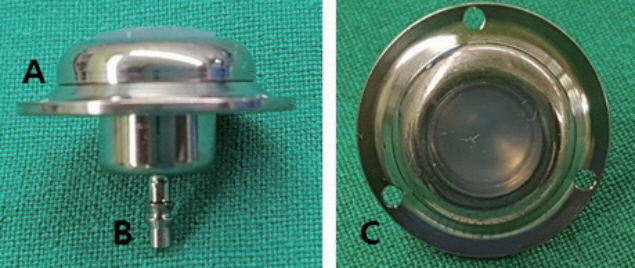
Fig. 1. Photographs of the V-Port. The V-Port is constituted of (A) a noncollapsible reservoir outlined with a titanium cage and (B) a connector for the ventricular catheter to be assembled. (C) The three holes of the titanium outer leaflet are designed to secure the V-Port on the skull using mini screws.

Fig. 2. Intraoperative photograph depicting V-Port insertion.
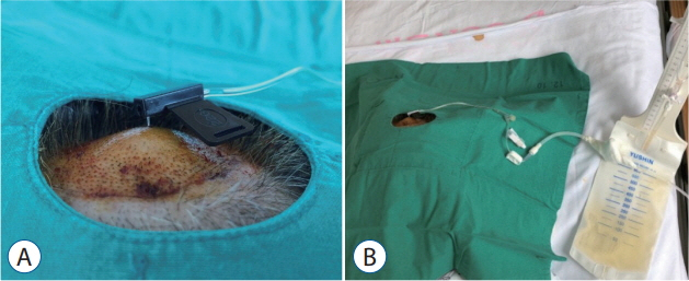
Fig. 3. Photographs of CSF being continuously drained. A : A hooked Huber needle was secured in the V-Port. B : The Huber needle was connected to an extra-ventricular drainage bag for continuous drainage of the CSF. CSF : cerebrospinal fluid.
Cited by 1 articles
-
Clinical Safety and Efficiency of the H-Port for Treatment of Leptomeningeal Metastasis
Sung-Min Jang, Ho-Shin Gwak, Ji-Woong Kwon, Sang Hoon Shin, Heon Yoo
J Korean Neurosurg Soc. 2024;67(4):467-476. doi: 10.3340/jkns.2023.0178.
Reference
-
References
1. Al-Anazi A, Bernstein M. Modified stereotactic insertion of the Ommaya reservoir. Technical note. J Neurosurg. 92:1050–1052. 2000.2. Gwak HS, Lee CH, Yang HS, Joo J, Shin SH, Yoo H, et al. Chemoport with a non-collapsible chamber as a replacement for an Ommaya reservoir in the treatment of leptomeningeal carcinomatosis. Acta Neurochir (Wien). 153:1971–1978. discussion 1978. 2011.
Article3. Hyun JW, Jeong IH, Joung A, Cho HJ, Kim SH, Kim HJ. Leptomeningeal metastasis: clinical experience of 519 cases. Eur J Cancer. 56:107–114. 2016.
Article4. Kak M, Nanda R, Ramsdale EE, Lukas RV. Treatment of leptomeningeal carcinomatosis: current challenges and future opportunities. J Clin Neurosci. 22:632–637. 2015.
Article5. Mack F, Baumert BG, Schäfer N, Hattingen E, Scheffler B, Herrlinger U, et al. Therapy of leptomeningeal metastasis in solid tumors. Cancer Treat Rev. 43:83–91. 2016.
Article6. Pace A, Fabi A. Chemotherapy in neoplastic meningitis. Crit Rev Oncol Hematol. 60:194–200. 2006.
Article7. Wang N, Bertalan MS, Brastianos PK. Leptomeningeal metastasis from systemic cancer: review and update on management. Cancer. 124:21–35. 2018.
Article
- Full Text Links
-
- Actions
-
Cited
- CITED
-
- Close
- Share
-
- Similar articles
-
- Traumatic Cerebrospinal Fluid Rhinorrhea: Successful Closure under the Surgical Microscope
- A Case of Spontaneous Intracranial Hypotension: Detection of Cerebrospinal Fluid Leakage by Early Dynamic Radionuclide Cisternography
- Endoscopic treatment of iatrogenic cerebrospinal fluid rhinorrhea
- Surgical management of iatrogenic cerebrospinal fluid rhinorrhea
- Traumatic Subdural Hygroma Due to Injury of an Ommaya Reservoir: A Case Report
