Metastatic Blue Nevus-Like Melanoma Detected by Liquid-Based Catheterized Urine Cytology
- Affiliations
-
- 1Department of Dermatology, Ajou University School of Medicine, Suwon, Korea.
- 2Department of Pathology, Ajou University School of Medicine, Suwon, Korea. kjefullup@naver.com
- KMID: 2419181
- DOI: http://doi.org/10.5021/ad.2018.30.3.356
Abstract
- Primary or metastatic malignant melanoma can mimic benign blue nevus in rare cases, making the diagnosis challenging. Herein, we report an exceptionally rare case of blue nevus-like melanoma and its blue nevus-like metastasis which was detected by catheterized urine cytology. The patient presented with blue-colored papuloplaques on his temple which were diagnosed as blue nevus-like melanoma on punch biopsies. While he was admitted for administration of chemotherapy, hematuria was detected. Catheterized urine cytology revealed singly scattered oval to spindle-shaped pigmented cells with a moderate degree of variation in shape and size. Many of them had small nuclei with indiscernible to inconspicuous nucleoli while only a few cells showed nuclear enlargement and nuclear hyperchromasia, which could be diagnostic pitfalls. Most of the cells on the smear were positive for HMB45 immunostaining, which confirmed the diagnosis of metastatic blue nevus-like melanoma. To the best of our knowledge, the present case is the first report describing cytomorphologic findings of blue nevus-like metastasis of melanoma in the urine specimen.
MeSH Terms
Figure
-

Fig. 1 A large ill-defined dark blue patch studded with pinhead to bean sized cobblestone surfaced papulonodules and satellite papules on the left temple.
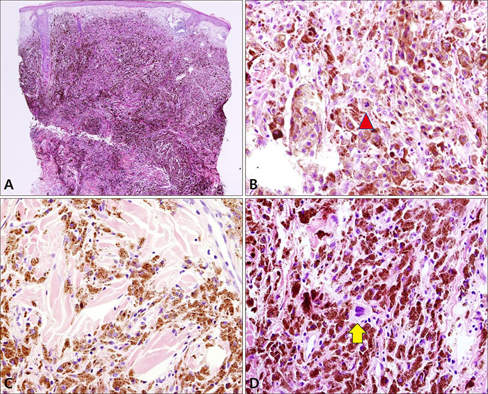
Fig. 2 Histologic figures of the specimen taken from the central (A, B) and peripheral (C, D) area of the lesion. (A) Solid proliferation of pigmented cells are noted in the entire dermis without junctional activity (H&E, ×40). (B) The tumor cells are composed of epithelioid and short spindle cells with mild to moderate nuclear enlargement, small nucleoli, and vesicular chromatin. A mitotic figure is identified in the deep dermis (arrowhead) (H&E, ×400). (C) The periphery of the lesion is composed of more spindle cells with minimal nuclear atypia (H&E, ×400). (D) Among the mildly atypical cells, there are rare bizarre atypical cells, visible on careful examination (arrow) (H&E, ×400).

Fig. 3 Catheterized urine cytology on liquid-based preparation. (A) The specimen is moderately cellular and composed of oval to spindle-shaped pigmented cells showing moderate degree of variations in size and shape, admixed with many red blood cells (papanicolaou [PAP] stain, virtual slide). (B) Many of the pigmented tumor cells have small nuclei, some of which are obscured by melanin pigment (arrowheads) (PAP, ×1,000). (C) A small number of cells shows increased nuclear to cytoplasmic ratio (arrow) (PAP, ×1,000). (D) Another atypical cell exhibit an enlarged angulated hyperchromatic nucleus (arrow) (PAP, ×1,000). (E) The pigmented cells are positive on HMB45 immunostaining (arrowheads) (amino ethyl carbazol [AEC], ×400).
Reference
-
1. Busam KJ. Metastatic melanoma to the skin simulating blue nevus. Am J Surg Pathol. 1999; 23:276–282.
Article2. Campa M, Patel M, Aubert P, Hosler G, Witheiler D. Blue nevus-like metastasis of a cutaneous melanoma identified by fluorescence in situ hybridization. Am J Dermatopathol. 2016; 38:695–697.
Article3. Granter SR, McKee PH, Calonje E, Mihm MC Jr, Busam K. Melanoma associated with blue nevus and melanoma mimicking cellular blue nevus: a clinicopathologic study of 10 cases on the spectrum of so-called ‘malignant blue nevus’. Am J Surg Pathol. 2001; 25:316–323.
Article4. Martin RC, Murali R, Scolyer RA, Fitzgerald P, Colman MH, Thompson JF. So-called “malignant blue nevus”: a clinicopathologic study of 23 patients. Cancer. 2009; 115:2949–2955.5. Costa S, Byrne M, Pissaloux D, Haddad V, Paindavoine S, Thomas L, et al. Melanomas associated with blue nevi or mimicking cellular blue nevi: clinical, pathologic, and molecular study of 11 cases displaying a high frequency of gna11 mutations, bap1 expression loss, and a predilection for the scalp. Am J Surg Pathol. 2016; 40:368–377.6. Loghavi S, Curry JL, Torres-Cabala CA, Ivan D, Patel KP, Mehrotra M, et al. Melanoma arising in association with blue nevus: a clinical and pathologic study of 24 cases and comprehensive review of the literature. Mod Pathol. 2014; 27:1468–1478.
Article7. Zogno C, Schiaffino E, Boeri R, Schmid C. Cytologic detection of metastatic malignant melanoma in urine. A report of three cases. Acta Cytol. 1997; 41:4 Suppl. 1332–1336.8. Meunier R, Pareek G, Amin A. Metastasis of malignant melanoma to urinary bladder: a case report and review of the literature. Case Rep Pathol. 2015; DOI: 10.1155/2015/173870.
Article9. Zembowicz A, Carney JA, Mihm MC. Pigmented epithelioid melanocytoma: a low-grade melanocytic tumor with metastatic potential indistinguishable from animal-type melanoma and epithelioid blue nevus. Am J Surg Pathol. 2004; 28:31–40.10. Urso C, Ginanneschi C, Anichini C, Paglierani M, Saieva C, Pimpinelli N, et al. Animal-type melanoma: report of five cases with sentinel node biopsy and fluorescence in-situ hybridization analysis. Melanoma Res. 2014; 24:47–53.11. Pouryazdanparast P, Newman M, Mafee M, Haghighat Z, Guitart J, Gerami P. Distinguishing epithelioid blue nevus from blue nevus-like cutaneous melanoma metastasis using fluorescence in situ hybridization. Am J Surg Pathol. 2009; 33:1396–1400.
Article
