Construction of an Electrocardiogram Database Including 12 Lead Waveforms
- Affiliations
-
- 1Department of Biomedical Informatics, Ajou University School of Medicine, Suwon, Korea. d.yoon.ajou@gmail.com
- 2Department of Cardiology, Ajou University School of Medicine, Suwon, Korea.
- 3Department of Biomedical Sciences, Ajou University Graduate School of Medicine, Suwon, Korea.
- KMID: 2418174
- DOI: http://doi.org/10.4258/hir.2018.24.3.242
Abstract
OBJECTIVES
Electrocardiogram (ECG) data are important for the study of cardiovascular disease and adverse drug reactions. Although the development of analytical techniques such as machine learning has improved our ability to extract useful information from ECGs, there is a lack of easily available ECG data for research purposes. We previously published an article on a database of ECG parameters and related clinical data (ECG-ViEW), which we have now updated with additional 12-lead waveform information.
METHODS
All ECGs stored in portable document format (PDF) were collected from a tertiary teaching hospital in Korea over a 23-year study period. We developed software which can extract all ECG parameters and waveform information from the ECG reports in PDF format and stored it in a database (meta data) and a text file (raw waveform).
RESULTS
Our database includes all parameters (ventricular rate, PR interval, QRS duration, QT/QTc interval, P-R-T axes, and interpretations) and 12-lead waveforms (for leads I, II, III, aVR, aVL, aVF, V1, V2, V3, V4, V5, and V6) from 1,039,550 ECGs (from 447,445 patients). Demographics, drug exposure data, diagnosis history, and laboratory test results (serum calcium, magnesium, and potassium levels) were also extracted from electronic medical records and linked to the ECG information.
CONCLUSIONS
Electrocardiogram information that includes 12 lead waveforms was extracted and transformed into a form that can be analyzed. The description and programming codes in this case report could be a reference for other researchers to build ECG databases using their own local ECG repository.
MeSH Terms
Figure
-
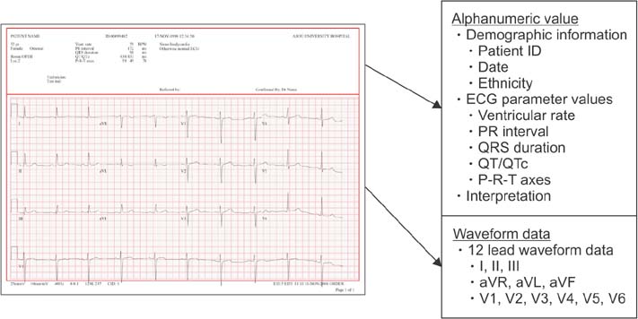
Figure 1 Example of an electrocardiogram (ECG) report. Alphanumeric values (demographic information, ECG parameter values, and interpretation) are located in the upper part of the report. Waveform data are given as time-series graphs with a grid, covering the middle and lower part of the report. One grid unit (1 mm × 1 mm square) corresponds to 0.1 mV × 0.04 seconds.

Figure 2 Data transformation process for the waveform data. (A) Raw data in SVG format contains information regarding the exact position of the data point on the electrocardiogram. (B) The start point of all 12-lead waveforms was set to 0. (C) Because 1 mV in the raw data corresponds to y-axis values of 28, we divided all y-axis values by 28 to adjust the scale of the y-axis to mV. (D) Using linear interpolation (500 Hz), vector image data given as x- and y-coordinates were converted into equidistant time series data. Although the resulting waveform data consist of a series of values without timestamps, the timestamps could be calculated by counting the data points from the starting point because the starting point was provided in the file name or database, and one data point corresponds to 1/500 seconds.
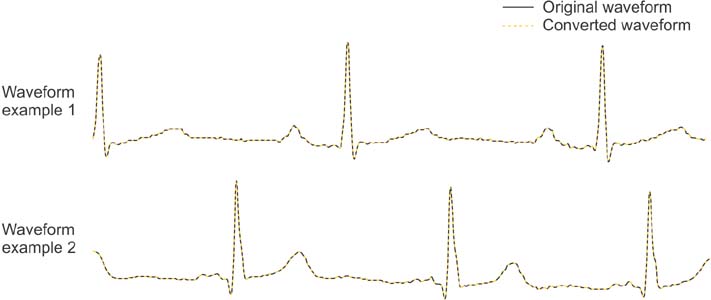
Figure 3 Comparison between the raw waveform and converted waveform data. Due to high-density interpolation, there was no significant difference between the raw data (which are based on x- and y-coordinates) and time series data.
Cited by 2 articles
-
Deep Learning-Based Electrocardiogram Signal Noise Detection and Screening Model
Dukyong Yoon, Hong Seok Lim, Kyoungwon Jung, Tae Young Kim, Sukhoon Lee
Healthc Inform Res. 2019;25(3):201-211. doi: 10.4258/hir.2019.25.3.201.Development of a Risk Score for QT Prolongation in the Intensive Care Unit Using Time-Series Electrocardiogram Data and Electronic Medical Records
Tae Young Kim, Byung Jin Choi, Yeryung Koo, Sukhoon Lee, Dukyong Yoon
Healthc Inform Res. 2021;27(3):182-188. doi: 10.4258/hir.2021.27.3.182.
Reference
-
1. Ovreiu M, Simon D. Biogeography-based optimization of neuro-fuzzy system parameters for diagnosis of cardiac disease. In : Proceedings of the 12th Annual Conference on Genetic and Evolutionary Computation; 2010 Jul 7–11; Portland, OR. p. 1235–1242.2. Martis RJ, Acharya UR, Adeli H. Current methods in electrocardiogram characterization. Comput Biol Med. 2014; 48:133–149.
Article3. O'Neil BJ, Hoekstra J, Pride YB, Lefebvre C, Diercks D, Frank Peacock W, et al. Incremental benefit of 80-lead electrocardiogram body surface mapping over the 12-lead electrocardiogram in the detection of acute coronary syndromes in patients without ST-elevation myocardial infarction: results from the Optimal Cardiovascular Diagnostic Evaluation Enabling Faster Treatment of Myocardial Infarction (OCCULT MI) trial. Acad Emerg Med. 2010; 17(9):932–939.4. Laguna P, Jane R, Caminal P. Automatic detection of wave boundaries in multilead ECG signals: validation with the CSE database. Comput Biomed Res. 1994; 27(1):45–60.
Article5. Lynch DR Jr, Washam JB, Newby LK. QT interval prolongation and torsades de pointes in a patient undergoing treatment with vorinostat: a case report and review of the literature. Cardiol J. 2012; 19(4):434–438.6. Li XB, Tang YL, Zheng W, Wang CY, de Leon J. QT interval prolongation associated with intramuscular ziprasidone in Chinese patients: a case report and a comprehensive literature review with meta-analysis. Case Rep Psychiatry. 2014; 2014:489493.
Article7. Tarapues M, Cereza G, Arellano AL, Montane E, Figueras A. Serious QT interval prolongation with ranolazine and amiodarone. Int J Cardiol. 2014; 172(1):e60–e61.8. Park MY, Yoon D, Choi NK, Lee J, Lee K, Lim HS, et al. Construction of an open-access QT database for detecting the proarrhythmia potential of marketed drugs: ECG-ViEW. Clin Pharmacol Ther. 2012; 92(3):393–396.
Article9. Kim YG, Shin D, Park MY, Lee S, Jeon MS, Yoon D, et al. ECG-ViEW II, a freely accessible electrocardiogram database. PLoS One. 2017; 12(4):e0176222.
Article10. Ortigosa N, Gimenez VM. Raw data extraction from electrocardiograms with Portable Document Format. Comput Methods Programs Biomed. 2014; 113(1):284–289.
Article11. Yoon D, Lee S, Kim TY, Ko J, Chung WY, Park RW. System for collecting biosignal data from multiple patient monitoring systems. Healthc Inform Res. 2017; 23(4):333–337.
Article
- Full Text Links
-
- Actions
-
Cited
- CITED
-
- Close
- Share
-
- Similar articles
-
- Occupational Diseases of Construction Industry
- A Case of Regressed Apical Hypertrophic Cardiomyopathy
- Changes of Brachial Arterial Doppler Waveform during Immersion of the Hand of Young Men in Ice-cold Water
- Source Data Bank Construction Of Nursing Research
- Standardized Database of 12-Lead Electrocardiograms with a Common Standard for the Promotion of Cardiovascular Research: KURIAS-ECG
