Is the diagnosis of calcified laryngeal cartilages on panoramic radiographs possible?
- Affiliations
-
- 1Department of Dentomaxillofacial Radiology, Faculty of Dentistry, Hacettepe University, Ankara, Turkey. lbartvinli@yahoo.com
- KMID: 2413923
- DOI: http://doi.org/10.5624/isd.2018.48.2.121
Abstract
- PURPOSE
Detecting laryngeal cartilages (triticeous and thyroid cartilages) on panoramic radiographs is important because they may be confused with carotid artery calcifications in the bifurcation region, which are a risk factor for stroke. This study assessed the efficiency of panoramic radiography in the diagnosis of calcified laryngeal cartilages using cone-beam computed tomography (CBCT) as the reference standard.
MATERIALS AND METHODS
A total of 312 regions (142 bilateral, 10 left, 18 right) in 170 patients (140 males, 30 females) were examined. Panoramic radiographs were examined by an oral and maxillofacial radiologist with 11 years of experience. CBCT scans were reviewed by 2 other oral and maxillofacial radiologists. The kappa coefficient (κ) was calculated to determine the level of intra-observer agreement and to determine the level of agreement between the 2 methods. Diagnostic indicators (sensitivity, specificity, accuracy, and false positive and false negative rates) were also calculated. P values < .05 were considered to indicate statistical significance.
RESULTS
Eighty-two images were re-examined to determine the intra-observer agreement level, and the kappa coefficient was calculated as 0.709 (P < .05). Statistically significant and acceptable agreement was found between the panoramic and CBCT images (κ=0.684 and P < .05). The sensitivity, specificity, diagnostic accuracy rate, the false positive rate, and the false negative rate of the panoramic radiographs were 85.4%, 83.5%, 84.6%, 16.5%, and 14.6%, respectively.
CONCLUSION
In most cases, calcified laryngeal cartilages could be diagnosed on panoramic radiographs. However, due to variation in the calcifications, diagnosis may be difficult.
Keyword
MeSH Terms
Figure
-
Fig. 1 A panoramic radiograph reveals small, ill-defined opacities below the greater horn of the hyoid on the right side, while calcification is demonstrated in the carotid region on cone-beam computed tomographic images.

Fig. 2 A panoramic radiograph reveals small, ill-defined opacities below the greater horn of the hyoid on the right side, while calcified laryngeal cartilages are demonstrated on cone-beam computed tomographic images.

Fig. 3 Axial maximum intensity projection of the patient in Figure 1 shows the location of the opacity. This image demonstrates calcification in the carotid region.
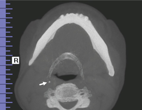
Fig. 4 Axial maximum intensity projection of the patient in Figure 2 shows the location of the opacity. This image demonstrates calcified laryngeal cartilages. Calcified laryngeal cartilages can be differentiated from calcification in the carotid region because they are medial to the greater horn of the hyoid bone.
Reference
-
1. Carter LC. Soft tissue calcifications and ossifications. In : White SC, Pharoah MJ, editors. Oral radiology: principles and interpretation. 7th ed. St. Louis: Elsevier;2014. p. 524–541.2. Kamikawa RS, Pereira MF, Fernandes A, Meurer MI. Study of the localization of radiopacities similar to calcified carotid atheroma by means of panoramic radiography. Oral Surg Oral Med Oral Pathol Oral Radiol Endod. 2006; 101:374–378.
Article3. Carter LC. Discrimination between calcified triticeous cartilage and calcified carotid atheroma on panoramic radiography. Oral Surg Oral Med Oral Pathol Oral Radiol Endod. 2000; 90:108–110.
Article4. Ahmad M, Madden R, Perez L. Triticeous cartilage: prevalence on panoramic radiographs and diagnostic criteria. Oral Surg Oral Med Oral Pathol Oral Radiol Endod. 2005; 99:225–230.
Article5. Mupparapu M, Vuppalapati A. Ossification of laryngeal cartilages on lateral cephalometric radiographs. Angle Orthod. 2005; 75:196–201.6. Scarfe WC, Li Z, Aboelmaaty W, Scott SA, Farman AG. Maxillofacial cone beam computed tomography: essence, elements and steps to interpretation. Aust Dent J. 2012; 57:Suppl 1. 46–60.
Article7. Alqahtani E, Marrero DE, Champion WL, Alawaji A, Kousoubris PD, Small JE. Triticeous cartilage CT imaging characteristics, prevalence, extent, and distribution of ossification. Otolaryngol Head Neck Surg. 2016; 154:131–137.
Article8. MacDonald D, Chan A, Harris A, Vertinsky T, Farman AG, Scarfe WC. Diagnosis and management of calcified carotid artery artheroma: dental perspectives. Oral Surg Oral Med Oral Pathol Oral Radiol. 2012; 114:533–547.9. Garoff M, Ahlqvist J, Levring Jäghagen E, Johansson E, Wester P. Study of the localization of radiopacities similar to calcified carotid atheroma by means of panoramic radiography. Carotid calcification in panoramic radiographs: radiographic appearance and the degree of carotid stenosis. (in press).
- Full Text Links
-
- Actions
-
Cited
- CITED
-
- Close
- Share
-
- Similar articles
-
- More frequent detection of calcified carotid atherosclerotic plaques and mineralized laryngeal cartilages on digital than on film-based panoramic radiographs
- Prediction of osteoporosis using fractal analysis on periapical and panoramic radiographs
- Comparison of panoramic radiography and cone-beam computed tomography for assessing radiographic signs indicating root protrusion into the maxillary sinus
- Projection angles of mandibular condyles in panoramic and transcranial radiographs
- Coincidence of calcified carotid atheromatous plaque, osteoporosis, and periodontal bone loss in dental panoramic radiographs
