J Vet Sci.
2017 Aug;18(S1):351-359. 10.4142/jvs.2017.18.S1.351.
Chimeric rabies glycoprotein with a transmembrane domain and cytoplasmic tail from Newcastle disease virus fusion protein incorporates into the Newcastle disease virion at reduced levels
- Affiliations
-
- 1Key Laboratory of Veterinary Public Health of Ministry of Agriculture, State Key Laboratory of Veterinary Biotechnology, Harbin Veterinary Research Institute, Chinese Academy of Agricultural Sciences, Harbin 150001, China. gjy2003@163.com
- KMID: 2412560
- DOI: http://doi.org/10.4142/jvs.2017.18.S1.351
Abstract
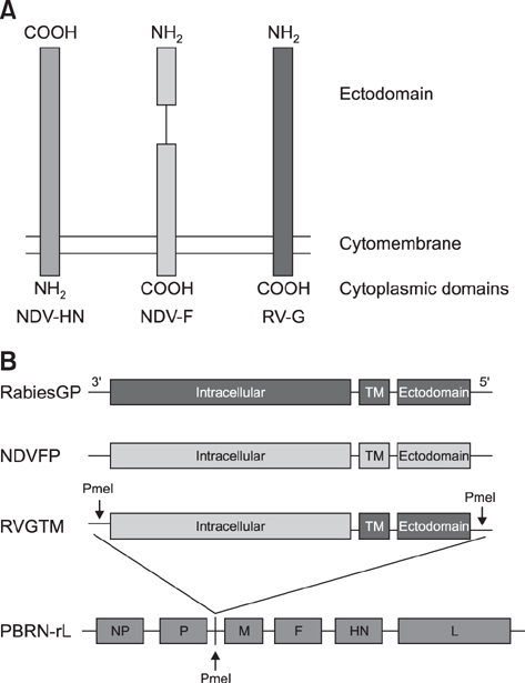
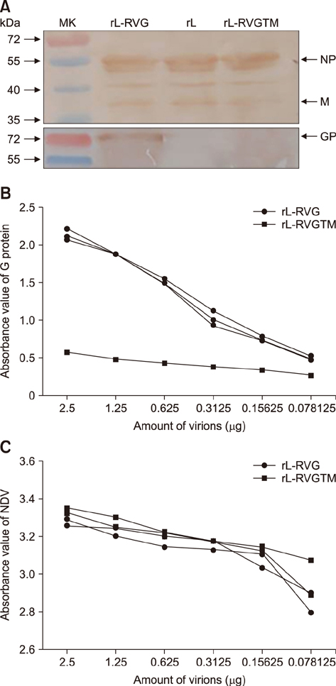
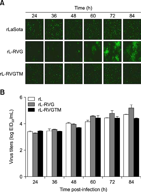
- Rabies remains an important worldwide health problem. Newcastle disease virus (NDV) was developed as a vaccine vector in animals by using a reverse genetics approach. Previously, our group generated a recombinant NDV (LaSota strain) expressing the complete rabies virus G protein (RVG), named rL-RVG. In this study, we constructed the variant rL-RVGTM, which expresses a chimeric rabies virus G protein (RVGTM) containing the ectodomain of RVG and the transmembrane domain (TM) and a cytoplasmic tail (CT) from the NDV fusion glycoprotein to study the function of RVG's TM and CT. The RVGTM did not detectably incorporate into NDV virions, though it was abundantly expressed at the surface of infected BHK-21 cells. Both rL-RVG and rL-RVGTM induced similar levels of NDV virus-neutralizing antibody (VNA) after initial and secondary vaccination in mice, whereas rabies VNA induction by rL-RVGTM was markedly lower than that induced by rL-RVG. Though rL-RVG could spread from cell to cell like that in rabies virus, rL-RVGTM lost this ability and spread in a manner similar to the parental NDV. Our data suggest that the TM and CT of RVG are essential for its incorporation into NDV virions and for spreading of the recombinant virus from the initially infected cells to surrounding cells.
Keyword
MeSH Terms
Figure
Reference
-
1. Albertini AA, Baquero E, Ferlin A, Gaudin Y. Molecular and cellular aspects of rhabdovirus entry. Viruses. 2012; 4:117–139.
Article2. Bukreyev A, Huang Z, Yang L, Elankumaran S, St Claire M, Murphy BR, Samal SK, Collins PL. Recombinant Newcastle disease virus expressing a foreign viral antigen is attenuated and highly immunogenic in primates. J Virol. 2005; 79:13275–13284.
Article3. Bukreyev A, Skiadopoulos MH, Murphy BR, Collins PL. Nonsegmented negative-strand viruses as vaccine vectors. J Virol. 2006; 80:10293–10306.
Article4. Chen L, Colman PM, Cosgrove LJ, Lawrence MC, Lawrence LJ, Tulloch PA, Gorman JJ. Cloning, expression, and crystallization of the fusion protein of Newcastle disease virus. Virology. 2001; 290:290–299.
Article5. Chen L, Gorman JJ, McKimm-Breschkin J, Lawrence LJ, Tulloch PA, Smith BJ, Colman PM, Lawrence MC. The structure of the fusion glycoprotein of Newcastle disease virus suggests a novel paradigm for the molecular mechanism of membrane fusion. Structure. 2001; 9:255–266.
Article6. Cleverley DZ, Lenard J. The transmembrane domain in viral fusion: essential role for a conserved glycine residue in vesicular stomatitis virus G protein. Proc Natl Acad Sci U S A. 1998; 95:3425–3430.
Article7. Della-Porta AJ, Spencer T. Newcastle disease. Aust Vet J. 1989; 66:424–426.
Article8. Dietzschold B, Schnell M, Koprowski H. Pathogenesis of rabies. Curr Top Microbiol Immunol. 2005; 292:45–56.
Article9. Dietzschold B, Wang HH, Rupprecht CE, Celis E, Tollis M, Ertl H, Heber-Katz E, Koprowski H. Induction of protective immunity against rabies by immunization with rabies virus ribonucleoprotein. Proc Natl Acad Sci U S A. 1987; 84:9165–9169.
Article10. Dietzschold B, Wiktor TJ, Trojanowski JQ, Macfarlan RI, Wunner WH, Torres-Anjel MJ, Koprowski H. Differences in cell-to-cell spread of pathogenic and apathogenic rabies virus in vivo and in vitro. J Virol. 1985; 56:12–18.
Article11. Dietzschold B, Wunner WH, Wiktor TJ, Lopes AD, Lafon M, Smith CL, Koprowski H. Characterization of an antigenic determinant of the glycoprotein that correlates with pathogenicity of rabies virus. Proc Natl Acad Sci U S A. 1983; 80:70–74.
Article12. DiNapoli JM, Kotelkin A, Yang L, Elankumaran S, Murphy BR, Samal SK, Collins PL, Bukreyev A. Newcastle disease virus, a host range-restricted virus, as a vaccine vector for intranasal immunization against emerging pathogens. Proc Natl Acad Sci U S A. 2007; 104:9788–9793.
Article13. DiNapoli JM, Ward JM, Cheng L, Yang L, Elankumaran S, Murphy BR, Samal SK, Collins PL, Bukreyev A. Delivery to the lower respiratory tract is required for effective immunization with Newcastle disease virus-vectored vaccines intended for humans. Vaccine. 2009; 27:1530–1539.
Article14. Doms RW, Keller DS, Helenius A, Balch WE. Role for adenosine triphosphate in regulating the assembly and transport of vesicular stomatitis virus G protein trimers. J Cell Biol. 1987; 105:1957–1969.
Article15. Faber M, Li J, Kean RB, Hooper DC, Alugupalli KR, Dietzschold B. Effective preexposure and postexposure prophylaxis of rabies with a highly attenuated recombinant rabies virus. Proc Natl Acad Sci U S A. 2009; 106:11300–11305.
Article16. Fu ZF. Rabies and rabies research: past, present and future. Vaccine. 1997; 15:Suppl. S20–S24.
Article17. Ge J, Deng G, Wen Z, Tian G, Wang Y, Shi J, Wang X, Li Y, Hu S, Jiang Y, Yang C, Yu K, Bu Z, Chen H. Newcastle disease virus-based live attenuated vaccine completely protects chickens and mice from lethal challenge of homologous and heterologous H5N1 avian influenza viruses. J Virol. 2007; 81:150–158.
Article18. Ge J, Tian G, Zeng X, Jiang Y, Chen H, Bua Z. Generation and evaluation of a Newcastle disease virus-based H9 avian influenza live vaccine. Avian Dis. 2010; 54:1 Suppl. S294–S296.
Article19. Ge J, Wang X, Tao L, Wen Z, Feng N, Yang S, Xia X, Yang C, Chen H, Bu Z. Newcastle disease virus-vectored rabies vaccine is safe, highly immunogenic, and provides long-lasting protection in dogs and cats. J Virol. 2011; 85:8241–8252.
Article20. Huang Z, Elankumaran S, Yunus AS, Samal SK. A recombinant Newcastle disease virus (NDV) expressing VP2 protein of infectious bursal disease virus (IBDV) protects against NDV and IBDV. J Virol. 2004; 78:10054–10063.
Article21. Jeetendra E, Ghosh K, Odell D, Li J, Ghosh HP, Whitt MA. The membrane-proximal region of vesicular stomatitis virus glycoprotein G ectodomain is critical for fusion and virus infectivity. J Virol. 2003; 77:12807–12818.
Article22. Khattar SK, Collins PL, Samal SK. Immunization of cattle with recombinant Newcastle disease virus expressing bovine herpesvirus-1 (BHV-1) glycoprotein D induces mucosal and serum antibody responses and provides partial protection against BHV-1. Vaccine. 2010; 28:3159–3170.
Article23. Maillard AP, Gaudin Y. Rabies virus glycoprotein can fold in two alternative, antigenically distinct conformations depending on membrane-anchor type. J Gen Virol. 2002; 83:1465–1476.
Article24. Morimoto K, Foley HD, McGettigan JP, Schnell MJ, Dietzschold B. Reinvestigation of the role of the rabies virus glycoprotein in viral pathogenesis using a reverse genetics approach. J Neurovirol. 2000; 6:373–381.
Article25. Morimoto K, Hooper DC, Spitsin S, Koprowski H, Dietzschold B. Pathogenicity of different rabies virus variants inversely correlates with apoptosis and rabies virus glycoprotein expression in infected primary neuron cultures. J Virol. 1999; 73:510–518.
Article26. Nakaya T, Cros J, Park MS, Nakaya Y, Zheng H, Sagrera A, Villar E, García-Sastre A, Palese P. Recombinant Newcastle disease virus as a vaccine vector. J Virol. 2001; 75:11868–11873.
Article27. Neumann G, Whitt MA, Kawaoka Y. A decade after the generation of a negative-sense RNA virus from cloned cDNA--what have we learned? J Gen Virol. 2002; 83:2635–2662.
Article28. Odell D, Wanas E, Yan J, Ghosh HP. Influence of membrane anchoring and cytoplasmic domains on the fusogenic activity of vesicular stomatitis virus glycoprotein G. J Virol. 1997; 71:7996–8000.
Article29. Park MS, Steel J, García-Sastre A, Swayne D, Palese P. Engineered viral vaccine constructs with dual specificity: avian influenza and Newcastle disease. Proc Natl Acad Sci U S A. 2006; 103:8203–8208.
Article30. Roche S, Gaudin Y. Characterization of the equilibrium between the native and fusion-inactive conformation of rabies virus glycoprotein indicates that the fusion complex is made of several trimers. Virology. 2002; 297:128–135.
Article31. Roche S, Rey FA, Gaudin Y, Bressanelli S. Structure of the prefusion form of the vesicular stomatitis virus glycoprotein G. Science. 2007; 315:843–848.
Article32. Veits J, Wiesner D, Fuchs W, Hoffmann B, Granzow H, Starick E, Mundt E, Schirrmeier H, Mebatsion T, Mettenleiter TC, Römer-Oberdörfer A. Newcastle disease virus expressing H5 hemagglutinin gene protects chickens against Newcastle disease and avian influenza. Proc Natl Acad Sci U S A. 2006; 103:8197–8202.
Article33. Weissenhorn W, Hinz A, Gaudin Y. Virus membrane fusion. FEBS Lett. 2007; 581:2150–2155.
Article34. Whitt MA, Buonocore L, Prehaud C, Rose JK. Membrane fusion activity, oligomerization, and assembly of the rabies virus glycoprotein. Virology. 1991; 185:681–688.
Article35. Wiktor TJ, Macfarlan RI, Reagan KJ, Dietzschold B, Curtis PJ, Wunner WH, Kieny MP, Lathe R, Lecocq JP, Mackett M. Protection from rabies by a vaccinia virus recombinant containing the rabies virus glycoprotein gene. Proc Natl Acad Sci U S A. 1984; 81:7194–7198.
Article36. Wyatt LS, Moss B, Rozenblatt S. Replication-deficient vaccinia virus encoding bacteriophage T7 RNA polymerase for transient gene expression in mammalian cells. Virology. 1995; 210:202–205.
Article
- Full Text Links
- Actions
-
Cited
- CITED
-
- Close
- Share
- Similar articles
-
- Expression of F Protein Gene of a Thermostable Isolate of Newcastle Disease Virus Using Baculovirus Expression System
- Antigenic and immunogenic investigation of the virulence motif of the Newcastle disease virus fusion protein
- Improved immunogenicity of Newcastle disease virus inactivated vaccine following DNA vaccination using Newcastle disease virus hemagglutinin-neuraminidase and fusion protein genes
- Red blood cell elution time of strains of Newcastle disease virus
- Newcastle disease virus vectored vaccines as bivalent or antigen delivery vaccines


