J Rheum Dis.
2018 Apr;25(2):140-143. 10.4078/jrd.2018.25.2.140.
Case of Polymyalgia Rheumatica Misdiagnosed as Infectious Spondylitis
- Affiliations
-
- 1Department of Internal Medicine, Hanyang University Seoul Hospital, Seoul, Korea. skchomd@hanyang.ac.kr
- 2Department of Rheumatology, Hanyang University Hospital for Rheumatic Diseases, Seoul, Korea.
- 3Department of Radiology, Hanyang University College of Medicine, Seoul, Korea.
- KMID: 2408932
- DOI: http://doi.org/10.4078/jrd.2018.25.2.140
Abstract
- A 60-year-old woman visited the authors' clinic with low back pain and arthralgia. Her symptoms had occurred 6 months previously, and she was treated with an epidural injection and a balloon dilatation procedure based on the assumption of spinal stenosis, but both treatments were ineffective. Her low back pain was aggravated, accompanied by fever and chills over a period of 4 months. As a result, she visited another referral hospital and was diagnosed with infective spondylitis associated with the invasive procedure. Her symptoms improved with antibiotics, but they recurred. When she visited our clinic, she still had continuous low back pain and febrile senses. Magnetic resonance imaging of her lumbar spine revealed interspinous bursitis, and 18 F-fluorodeoxyglucose positron emission tomography showed multifocal synovial inflammation. She was diagnosed with polymyalgia rheumatica and treatment was started on prednisolone and celecoxib. Her symptoms improved dramatically and the inflammatory markers normalized.
Keyword
MeSH Terms
-
Anti-Bacterial Agents
Arthralgia
Back Pain
Bursitis
Celecoxib
Chills
Dilatation
Female
Fever
Humans
Inflammation
Injections, Epidural
Low Back Pain
Magnetic Resonance Imaging
Middle Aged
Polymyalgia Rheumatica*
Positron-Emission Tomography
Prednisolone
Referral and Consultation
Spinal Stenosis
Spine
Spondylitis*
Anti-Bacterial Agents
Celecoxib
Prednisolone
Figure
-
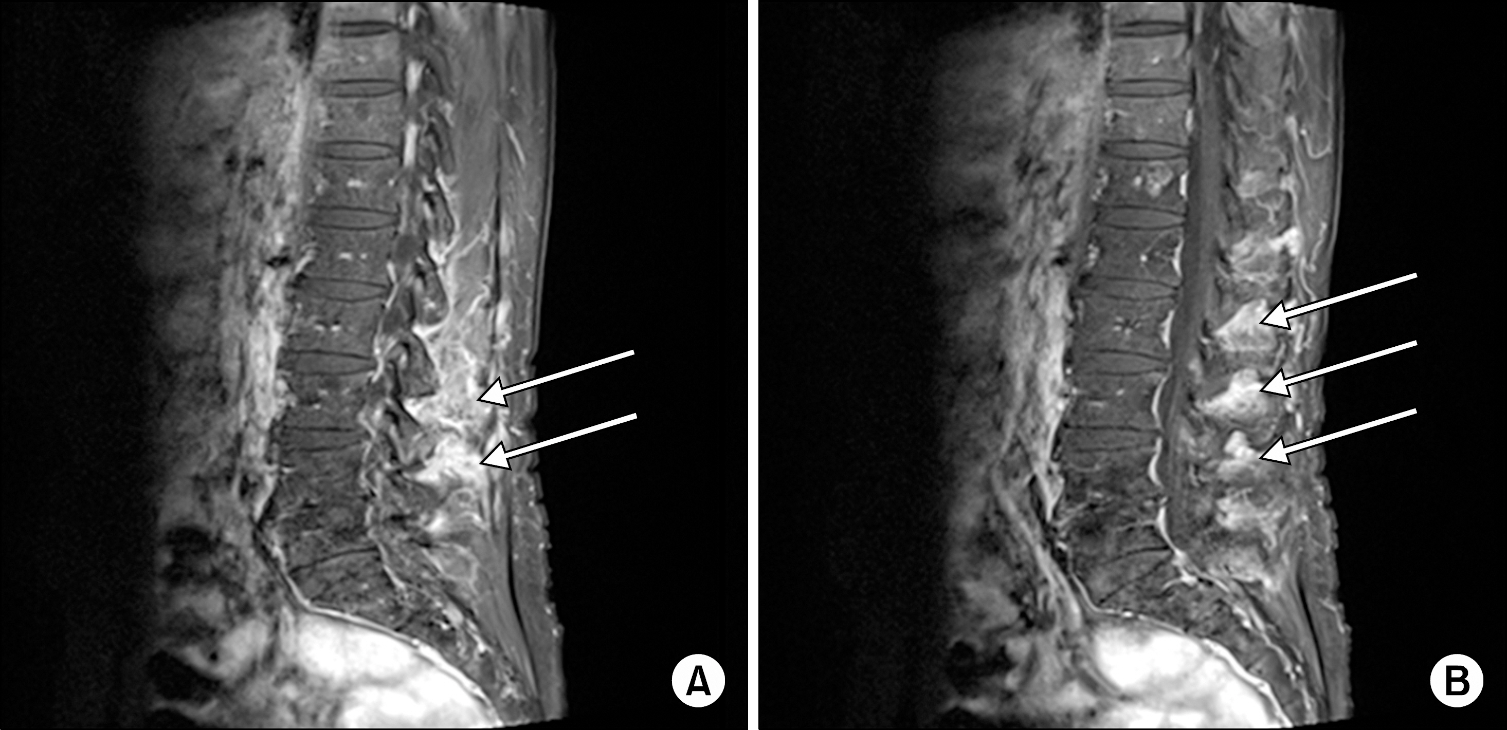
Figure 1. L-spine magnetic resonance imaging. (A) Diffuse enhancement along capsule and adjacent soft tissue at bilateral facet joint of L1-L5, (B) interspinous bursitis at T12-L5 on T1 weighted image.
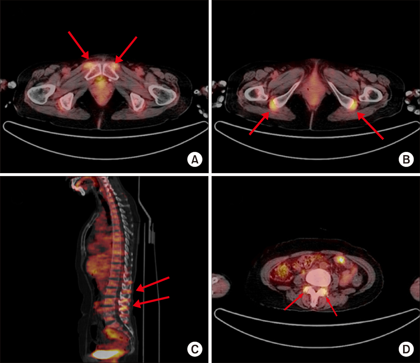
Figure 2. Whole body 18-fluo-rodeoxyglucose (FDG) positron emission tomography integrated with computed tomography scan. (A) Multiple inflammatory bursitis in pubic symphysis and (B) both ischiogluteal, trochan-teric bursa were revealed. In addition, increased FDG uptakes were noted at the (C) C6-7, T12-L5 level interspinous ligament and arthritis (D) on facet joints of L2-3 and L3-4 was revealed.
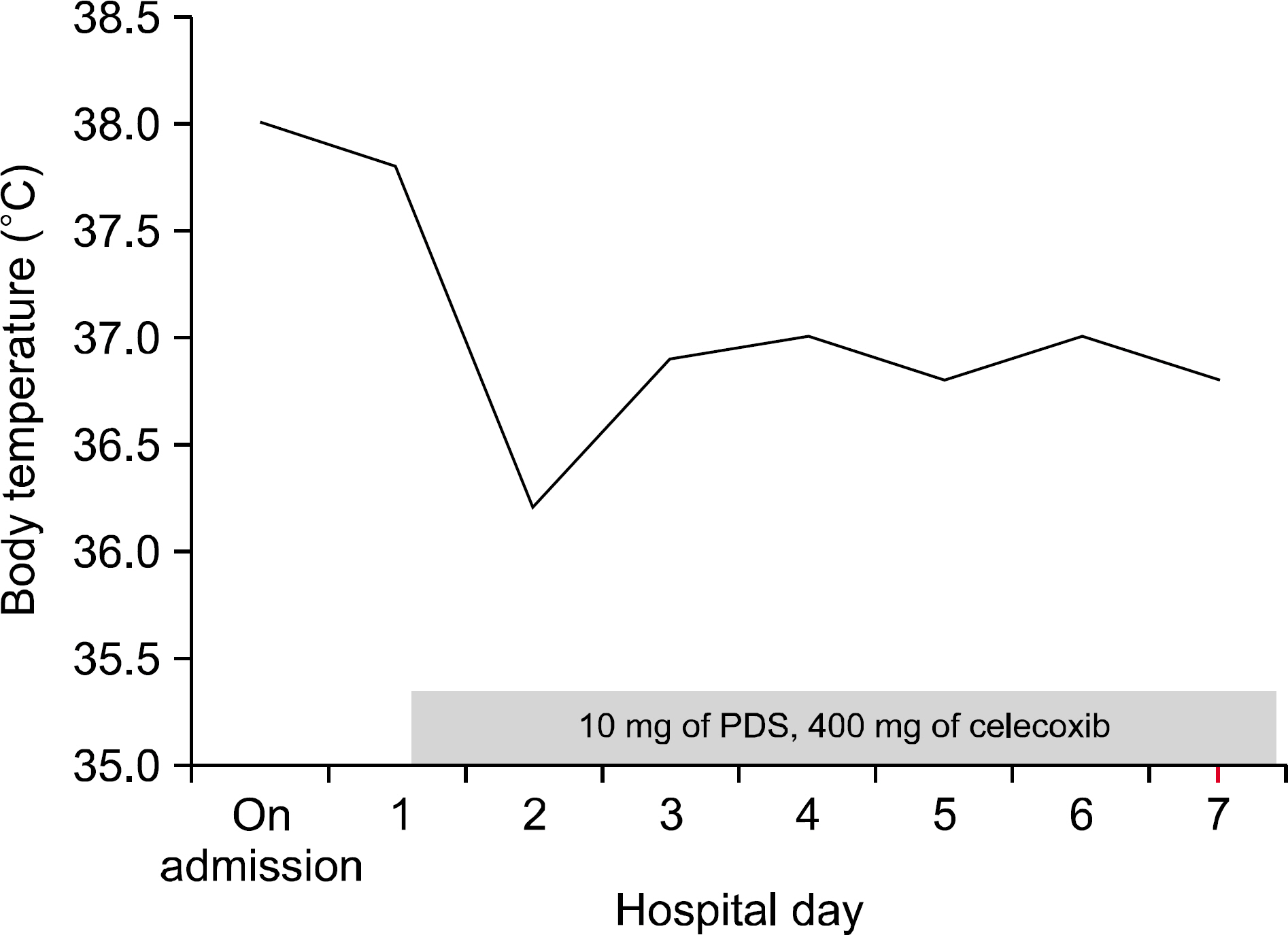
Figure 3. Clinical course of patient. PDS: prednisolone.
Reference
-
1. Gonzalez-Gay MA, Vazquez-Rodriguez TR, Lopez-Diaz MJ, Miranda-Filloy JA, Gonzalez-Juanatey C, Martin J, et al. Epidemiology of giant cell arteritis and polymyalgia rheumatica. Arthritis Rheum. 2009; 61:1454–61.
Article2. Kim IY, Seo GH, Lee S, Jeong H, Kim H, Lee J, et al. Epidemiology of polymyalgia rheumatica in Korea. J Rheum Dis. 2014; 21:297–302.
Article3. Salvarani C, Pipitone N, Versari A, Hunder GG. Clinical features of polymyalgia rheumatica and giant cell arteritis. Nat Rev Rheumatol. 2012; 8:509–21.
Article4. Dasgupta B, Cimmino MA, Kremers HM, Schmidt WA, Schirmer M, Salvarani C, et al. 2012 Provisional classification criteria for polymyalgia rheumatica: a European League Against Rheumatism/American College of Rheumatology collaborative initiative. Arthritis Rheum. 2012; 64:943–54.
Article5. Camellino D, Cimmino MA. Imaging of polymyalgia rheumatica: indications on its pathogenesis, diagnosis and prognosis. Rheumatology (Oxford). 2012; 51:77–86.
Article6. Park JS, Pyo JY, Park HJ, Lee HS, Kang Y, Kang MI, et al. Typical 18-FDG-PET/CT findings of polymyalgia rheumatica: a case report. J Rheum Dis. 2013; 20:113–7.
Article7. Camellino D, Paparo F, Morbelli S, Cutolo M, Sambuceti G, Cimmino MA. Interspinous bursitis is common in polymyalgia rheumatica, but is not associated with spinal pain. Arthritis Res Ther. 2014; 16:492.
Article8. Yamashita H, Kubota K, Takahashi Y, Minaminoto R, Morooka M, Ito K, et al. Whole-body fluorodeoxyglucose positron emission tomography/computed tomography in patients with active polymyalgia rheumatica: evidence for distinctive bursitis and large-vessel vasculitis. Mod Rheumatol. 2012; 22:705–11.
Article9. Salvarani C, Barozzi L, Boiardi L, Pipitone N, Bajocchi GL, Macchioni PL, et al. Lumbar interspinous bursitis in active polymyalgia rheumatica. Clin Exp Rheumatol. 2013; 31:526–31.10. Sondag M, Guillot X, Verhoeven F, Blagosklonov O, Prati C, Boulahdour H, et al. Utility of 18F-fluoro-dexoxyglucose positron emission tomography for the diagnosis of polymyalgia rheumatica: a controlled study. Rheumatology (Oxford). 2016; 55:1452–7.
Article11. Wendling D, Blagosklonov O, Boulahdour H, Prati C. Positron emission tomography: the ideal tool in polymyalgia rheumatica? Joint Bone Spine. 2014; 81:381–3.
Article
- Full Text Links
-
- Actions
-
Cited
- CITED
-
- Close
- Share
-
- Similar articles
-
- A Case of polymyalgia rheumatica
- Treatment Experience with Polymyalgia Rheumatica: A report of two cases
- A Case of Amyloidosis Presenting as Chronic Cholecystitis, Misdiagnosed as Polymyalgia Rheumatica
- Localized Mesenteric Vasculitis in a Patient with Polymyalgia Rheumatica
- Korean Epidemiologic Study of Polymyalgia Rheumatica
