Straight-Forward versus Bicortical Fixation Penetrating Endplate in Lumbosacral Fixation-A Biomechanical Study
- Affiliations
-
- 1Department of Orthopaedics and Traumatology, Dokuz Eylul University, Faculty of Medicine, Izmir, Turkey.
- 2Department of Orthopaedics and Traumatology, Catalca Ilyas Cokay Hospital, Istanbul, Turkey. zenanacar@gmail.com
- 3Department of Biomechanics, Dokuz Eylul University, Health Science Institute, Izmir, Turkey.
- KMID: 2408018
- DOI: http://doi.org/10.3340/jkns.2017.0404.004
Abstract
OBJECTIVE
Many lumbosacral fixation techniques have been described to offer a more screw-bone purchase. The forward anatomical fixation parallel to the endplate is still the most preferred method. Literature revealed little knowledge regarding the mechanical stability of lumbosacral trans-endplate fixation compared to the traditional trans-pedicular screw fixation method. The aim of this study is to assess the pull-out strength of lumbosacral screws penetrating the end plate and comparing it to the conventional trans-pedicular screw insertion method.
METHODS
Eight lumbar and eight sacral vertebrae, with average age 69.4 years, Left pedicles of the 5th lumbar vertebrae were used for trans-endplate screw fixation, group 1A, right pedicles were used for anatomical trans-pedicular screw fixation, group 1B. In the sacral vertebrae, the right side S1 pedicles were used for trans-endplate fixation, group 2A, left side pedicles were used for anatomical trans-pedicular screw fixation, group 2B. The biomechanical tests were performed using the axial compression testing machine. All tests were applied using 2 mm/min traction speed.
RESULTS
The average pull-out strength values of groups 1A and 1B were 403.78±11.71 N and 306.26±17.55 N, respectively. A statistical significance was detected with p=0.012. The average pull-out strength values of groups 2A and 2B were 388.73±17.03 N and 299.84±17.52 N, respectively. A statistical significance was detected with p=0.012.
CONCLUSION
The trans-endplate lumbosacral fixation method is a trustable fixation method with a stronger screw-bone purchase and offer a good alternative for surgeons specially in patients with osteoporosis.
Keyword
Figure
-
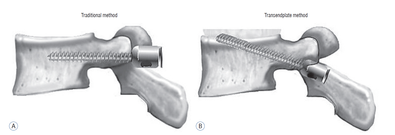
Fig. 1 A : Traditional trans-pedicular screw fixation through the body parallel to the end plate. B : Trans-endplate screw fixation penetrating the upper end plate, starting from the level of the inferior border of the transverse process, just 2 mm distal to the pedicle entrance, the trans-endplate lumbar pedicle screw is introduced 30° cranially to penetrate the superolateral anterior aspect of the end plate.
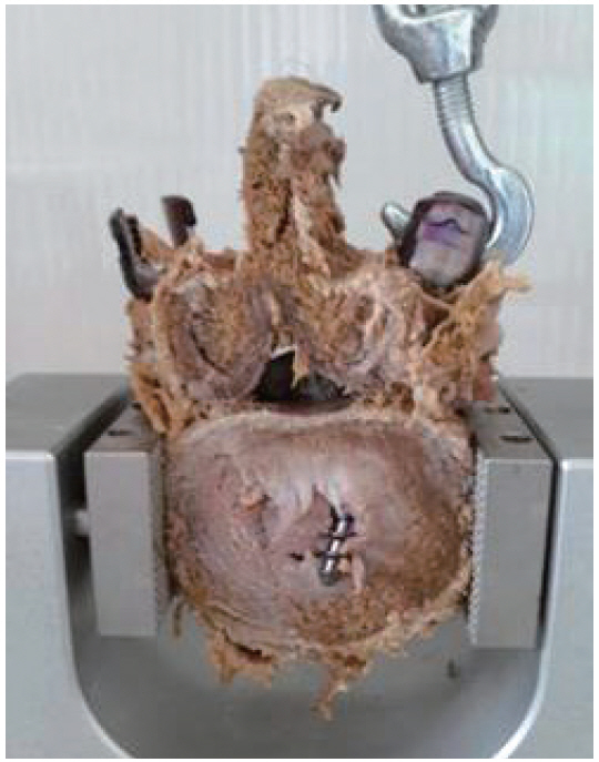
Fig. 2 The left pedicles of the 5th lumbar vertebrae were used for transendplate screw fixation penetrating the upper end plate, 2 mm distal to the pedicle entrance the lumbar pedicle screw was introduced 30° cranially to penetrate the superolateral and anterior aspect of the end plate. The right sides of the 5th lumbar vertebrae were used for anatomical transpedicular screw fixation through the body parallel to the end plate.
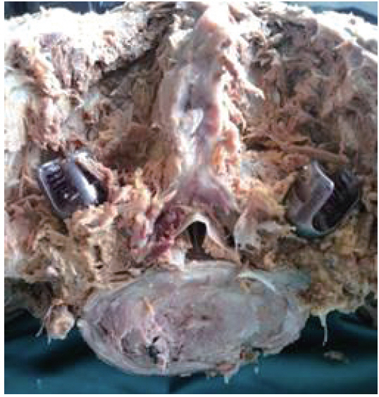
Fig. 3 The right sides of S1 pedicles were used for trans-endplate fixation penetrating S1 upper end plate, just 2 mm distal to the pedicle entrance the screw was directed cranially 30° to penetrate the superolateral and anterior aspect of the upper end plate of S1. The left side pedicles of S1 vertebra were used for anatomical trans-pedicular screw fixation through the body of S1 without penetrating the anterior cortex.
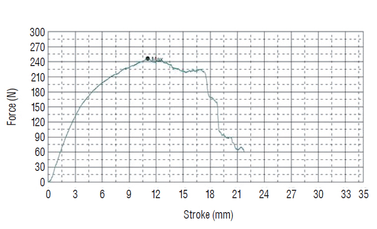
Fig. 4 The biomechanical tests were performed using the axial compression testing machine (AG-I 10 kN, Shimadzu Shikenki Engineering Co., Ltd, Shimadzu, Japan). All tests were applied using 2 mm/min traction speed.
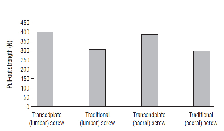
Fig. 5 A bar graph demonstrating the pull-out strength differences between different fixation techniques in L5 and S1 vertebrae.
Reference
-
References
1. Acar N, Karakasli A, Karaarslan AA, Ozcanhan MH, Ertem F, Erduran M. The mechanical effect of rod contouring on rod-screw system strength in spine fixation. J Korean Neurosurg Soc. 59:425–429. 2016.
Article2. Alkaly RN, Bader DL. The effect of transpedicular screw design on its performance in vertebral bone under tensile loads: a parametric study. Clin Spine Surg. 29:433–440. 2016.
Article3. Balderston RA, Winter RB, Moe JH, Bradford DS, Lonstein JE. Fusion to the sacrum for nonparalytic scoliosis in the adult. Spine (Phila Pa 1976). 11:824–829. 1986.
Article4. Carlson GD, Abitbol JJ, Anderson DR, Krag MH, Kostuik JP, Woo SL, et al. Screw fixation in the human sacrum. An in vitro study of the biomechanics of fixation. Spine (Phila Pa 1976). 17(6 Suppl):S196–S203. 1992.5. Cook SD, Salkeld SL, Stanley T, Faciane A, Miller SD. Biomechanical study of pedicle screw fixation in severely osteoporotic bone. Spine J. 4:402–408. 2004.
Article6. Cunningham BW, Sefter JC, Shono Y, McAfee PC. Static and cyclical biomechanical analysis of pedicle screw spinal constructs. Spine (Phila Pa 1976). 18:1677–1688. 1993.
Article7. Esses SI, Botsford DJ, Huler RJ, Rauschning W. Surgical anatomy of the sacrum. A guide for rational screw fixation. Spine (Phila Pa 1976). 16(6 Suppl):S283–S288. 1991.8. Gazzeri R. Percutaneous pedicle screw fixation technique in the thoracic and lumbar spine-tips and tricks. Surg Technol Int. 28:303–310. 2016.9. Hirano T, Hasegawa K, Takahashi HE, Uchiyama S, Hara T, Washio T, et al. Structural characteristic f the pedicle and its role in screw stability. Spine (Phila Pa 1976). 22:2504–2509. discussion 2510. 1997.10. Jiang L, Arlet V, Beckman L, Steffen T. Double pedicle screw instrumentation in the osteoporotic spine : a biomechanical feasibility study. J Spinal Disord Tech. 20:430–435. 2007.11. Jackson RP, Hamilton AC. CD screws with oblique canals for improved sacral fixation: a prospective clinical study of the first fifty patients. In : 7th proceeding of the International Congress on Cotrel-Dubousset Instrumentation; Montpellier: Sauramps Medical;1990. p. 75–86.12. Karakaşlı A, Sekik E, Karaarslan A, Kızmazoğlu C, Havıtçıoğlu H. Are pedicular screws and lateral hook screws more resistant against pullout than conventional spinal hooks and screws in terminal vertebral segment fixation? Eklem Hastalik Cerrahisi. 27:22–28. 2016.
Article13. Luk KD, Chen L, Lu WW. A stronger bicortical sacral pedicle screw fixation through the S1 endplate: an In vitro cyclic loading and pull-out force evaluation. Spine (Phila Pa 1976). 30:525–529. 2005.
Article14. Kettler A, Schmoelz W, Shezifi Y, Ohana N, Ben-Arye A, Claes L, et al. Biomechanical performance of the new beadex implant in the treatment of osteoporotic vertebral body compression fractures: restoration and maintenance of height and stability. Clin Biomech (Bristol, Avon). 21:676–682. 2006.
Article15. Krag MH. Biomechanics of transpedicle spinal fixation. In : Weinstein JN, Wiesel S, editors. The Lumbar Spine. Philadelphia: WBSaunders;1990. p. 916–940.16. Krag MH. Lumbosacral fixation with the vermont spinal fixator. In : Lin PM, Gill K, editors. Lumbar Interbody Fusion: Principles and Techniques of Spine Surgery. Rockville: Aspen Public;1988. p. 251–260.17. Leong JC, Lu WW, Zheng Y, Zhu Q, Zhong S. Comparison of the strengths of lumbosacral fixation achieved with techniques using one and two triangulated sacral screws. Spine (Phila Pa 1976). 23:2289–2294. 1998.
Article18. Licht NJ, Rowe DE, Ross LM. Pitfalls of pedicle screw fixation in the sacrum. A cadaver model. Spine (Phila Pa 1976). 17:892–896. 1992.19. Matsukawa K, Yato Y, Kato T, Imabayashi H, Asazuma T, Nemoto K. Cortical bone trajectory for lumbosacral fixation: penetrating S-1 endplate screw technique: technical note. J Neurosurg Spine. 21:203–209. 2014.
Article20. Mirkovic S, Abitol JJ, Steinman J, Edwards CC, Schaffler M, Massie J, et al. Anatomic consideration for sacral screw placement. Spine (Phila Pa 1976). 16(6 Suppl):S289–S294. 1991.
Article21. Ogilvie JW, Schendel M. Comparison of lumbosacral fixation devices. Clin Orthop Relat Res. (203):120–125. 1986.
Article22. Renner SM, Lim TH, Kim WJ, Katolik L, An HS, Andersson GB. Augmentation of pedicle screw fixation strength using an injectable calcium phosphate cement as a function of injection timing and method. Spine (Phila Pa 1976). 29:E212–E216. 2004.
Article23. Zheng Y, Lu WW, Zhu Q, Qin L, Zhong S, Leong JC. Variation in bone mineral density of the sacrum in young adults and its significance for sacral fixation. Spine (Phila Pa 1976). 25:353–357. 2000.
Article
- Full Text Links
-
- Actions
-
Cited
- CITED
-
- Close
- Share
-
- Similar articles
-
- The Comparison of LC-DCP versus LCP Fixation in the Plate Augmentation for the Nonunion of Femur Shaft Fractures after Intramedullary Nail Fixation
- Anatomical Safe Zone of Sacral Ala for Ventrolateral Sacral(S1) Screw Placement: Re-evaluation of Its Effectiveness
- Spinopelvic Fixation
- Is S1 Alar Iliac Screw a Feasible Option for Lumbosacral Fixation?: A Technical Note
- Comparison of the Fixation Strengths of Screws between the Traditional Trajectory and the Single and Double Endplate Penetrating Screw Trajectories Using Osteoporotic Vertebral Body Models Based on the Finite Element Method
