Neuroradiological and Neurophysiological Characteristics of Patients With Dyskinetic Cerebral Palsy
- Affiliations
-
- 1Department of Physical Medicine and Rehabilitation, Chonbuk National University Medical School, Jeonju, Korea. shpark0130@chonbuk.ac.kr
- 2Department of Radiology, Chonbuk National University Medical School, Jeonju, Korea.
Abstract
OBJECTIVE
To investigate neuroradiological and neurophysiological characteristics of patients with dyskinetic cerebral palsy (CP), by using magnetic resonance imaging (MRI), voxel-based morphometry (VBM), diffusion tensor tractography (DTT), and motor evoked potential (MEP).
METHODS
Twenty-three patients with dyskinetic CP (13 males, 10 females; mean age 34 years, range 16-50 years) were participated in this study. Functional evaluation was assessed by the Gross Motor Functional Classification System (GMFCS) and Barry-Albright Dystonia Scale (BADS). Brain imaging was performed on 3.0 Tesla MRI, and volume change of the grey matter was assessed using VBM. The corticospinal tract (CST) and superior longitudinal fasciculus (SLF) were analyzed by DTT. MEPs were recorded in the first dorsal interossei, the biceps brachii and the deltoid muscles.
RESULTS
Mean BADS was 16.4+/-5.0 in ambulatory group (GMFCS levels I, II, and III; n=11) and 21.3+/-3.9 in non-ambulatory group (GMFCS levels IV and V; n=12). Twelve patients showed normal MRI findings, and eleven patients showed abnormal MRI findings (grade I, n=5; grade II, n=2; grade III, n=4). About half of patients with dyskinetic CP showed putamen and thalamus lesions on MRI. Mean BADS was 20.3+/-5.7 in normal MRI group and 17.5+/-4.0 in abnormal MRI group. VBM showed reduced volume of the hippocampus and parahippocampal gyrus. In DTT, no abnormality was observed in CST, but not in SLF. In MEPs, most patients showed normal central motor conduction time.
CONCLUSION
These results support that extrapyramidal tract, related with basal ganglia circuitry, may be responsible for the pathophysiology of dyskinetic CP rather than CST abnormality.
Keyword
MeSH Terms
Figure
-

Fig. 1 There was a significant difference in Barry-Albright Dystonia Scale (BADS) scores between the ambulatory group and non-ambulatory group. *p<0.05.
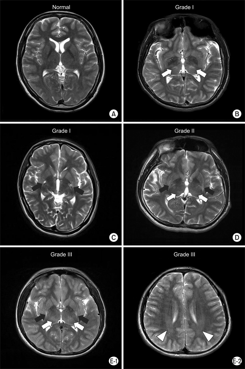
Fig. 2 Axial T2-weighted image. (A) No evidence of abnormality in patient no. 9. (B) Bilateral focal hyperintensities in the thalamus (white arrows) in patient no. 19. (C) Bilateral focal hyperintensities in the putamen (black arrows) in patient no. 17. (D) Bilateral focal hyperintensities in the putamen (black arrows) and thalamus (white arrows) in patient no. 21. (E-1) Bilateral focal hyperintensities in the putamen (black arrows) and thalamus (white arrows) in patient no. 1. (E-2) Bilateral diffuse hyperintensities in the periventricular white matter (white arrow heads) in patient no. 1.
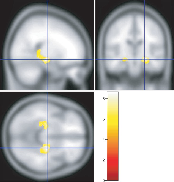
Fig. 3 Voxel-based morphometry analysis showed the regions with significantly reduced grey matter volumes in patients with dyskinetic cerebral palsy in yellow color (p<0.05, corrected for multiple comparison, familywise error rate).
Reference
-
1. Christine C, Dolk H, Platt MJ, Colver A, Prasauskiene A, Krageloh-Mann I, et al. Recommendations from the SCPE collaborative group for defining and classifying cerebral palsy. Dev Med Child Neurol Suppl. 2007; 109:35–38. PMID: 17370480.2. Himmelmann K, Hagberg G, Wiklund LM, Eek MN, Uvebrant P. Dyskinetic cerebral palsy: a population-based study of children born between 1991 and 1998. Dev Med Child Neurol. 2007; 49:246–251. PMID: 17376133.
Article3. Himmelmann K, McManus V, Hagberg G, Uvebrant P, Krageloh-Mann I, Cans C, et al. Dyskinetic cerebral palsy in Europe: trends in prevalence and severity. Arch Dis Child. 2009; 94:921–926. PMID: 19465585.
Article4. Andersen GL, Irgens LM, Haagaas I, Skranes JS, Meberg AE, Vik T. Cerebral palsy in Norway: prevalence, subtypes and severity. Eur J Paediatr Neurol. 2008; 12:4–13. PMID: 17574886.5. Barry MJ, VanSwearingen JM, Albright AL. Reliability and responsiveness of the Barry-Albright Dystonia Scale. Dev Med Child Neurol. 1999; 41:404–411. PMID: 10400175.
Article6. Monbaliu E, Ortibus E, Roelens F, Desloovere K, Deklerck J, Prinzie P, et al. Rating scales for dystonia in cerebral palsy: reliability and validity. Dev Med Child Neurol. 2010; 52:570–575. PMID: 20132143.
Article7. Ashwal S, Russman BS, Blasco PA, Miller G, Sandler A, Shevell M, et al. Practice parameter: diagnostic assessment of the child with cerebral palsy: report of the Quality Standards Subcommittee of the American Academy of Neurology and the Practice Committee of the Child Neurology Society. Neurology. 2004; 62:851–863. PMID: 15037681.
Article8. Seo JS, Kim TM, Chae JM, Kim YK, Kim BO. Comparative analysis of developmental assessment, evoked potentials, electroencephalography, and brain MRI in children with cerebral palsy. J Korean Acad Rehabil Med. 2000; 24:645–656.9. Park CI, Kim SW, Kim YC, Shin JC, Lee JD. Brain MRI and SPECT findings in children with cerebral palsy. J Korean Acad Rehabil Med. 1997; 21:1060–1067.10. Krageloh-Mann I, Helber A, Mader I, Staudt M, Wolff M, Groenendaal F, et al. Bilateral lesions of thalamus and basal ganglia: origin and outcome. Dev Med Child Neurol. 2002; 44:477–484. PMID: 12162385.
Article11. Bax M, Tydeman C, Flodmark O. Clinical and MRI correlates of cerebral palsy: the European Cerebral Palsy Study. JAMA. 2006; 296:1602–1608. PMID: 17018805.12. Einspieler C, Cioni G, Paolicelli PB, Bos AF, Dressler A, Ferrari F, et al. The early markers for later dyskinetic cerebral palsy are different from those for spastic cerebral palsy. Neuropediatrics. 2002; 33:73–78. PMID: 12075487.
Article13. Baron JC, Chetelat G, Desgranges B, Perchey G, Landeau B, de la Sayette V, et al. In vivo mapping of gray matter loss with voxel-based morphometry in mild Alzheimer's disease. Neuroimage. 2001; 14:298–309. PMID: 11467904.
Article14. Muller K, Homberg V, Aulich A, Lenard HG. Magnetoelectrical stimulation of motor cortex in children with motor disturbances. Electroencephalogr Clin Neurophysiol. 1992; 85:86–94. PMID: 1373370.15. Yokochi K, Aiba K, Kodama M, Fujimoto S. Magnetic resonance imaging in athetotic cerebral palsied children. Acta Paediatr Scand. 1991; 80:818–823. PMID: 1957601.
Article16. Good CD, Johnsrude IS, Ashburner J, Henson RN, Friston KJ, Frackowiak RS. A voxel-based morphometric study of ageing in 465 normal adult human brains. Neuroimage. 2001; 14(1 Pt 1):21–36. PMID: 11525331.
Article17. Mori S, Crain BJ, Chacko VP, van Zijl PC. Three-dimensional tracking of axonal projections in the brain by magnetic resonance imaging. Ann Neurol. 1999; 45:265–269. PMID: 9989633.
Article18. Vernooij MW, Smits M, Wielopolski PA, Houston GC, Krestin GP, van der Lugt A. Fiber density asymmetry of the arcuate fasciculus in relation to functional hemispheric language lateralization in both right- and left-handed healthy subjects: a combined fMRI and DTI study. Neuroimage. 2007; 35:1064–1076. PMID: 17320414.
Article19. Dominkus M, Grisold W, Jelinek V. Transcranial electrical motor evoked potentials as a prognostic indicator for motor recovery in stroke patients. J Neurol Neurosurg Psychiatry. 1990; 53:745–748. PMID: 2174076.
Article20. Yook SW, Park SH, Ko MH, Seo JH. Motor evoked potentials of the upper extremities in healthy children. Ann Rehabil Med. 2011; 35:759–764. PMID: 22506203.
Article21. Worsley KJ, Marrett S, Neelin P, Vandal AC, Friston KJ, Evans AC. A unified statistical approach for determining significant signals in images of cerebral activation. Hum Brain Mapp. 1996; 4:58–73. PMID: 20408186.
Article22. Kretzschmar K, Ludwig B, Kramer G, Collmann H, Kazner E. Bilateral lesions of the putamina. Neuroradiology. 1986; 28:87–91. PMID: 3951696.
Article23. Krageloh-Mann I. Imaging of early brain injury and cortical plasticity. Exp Neurol. 2004; 190(Suppl 1):S84–S90. PMID: 15498546.24. Maegaki Y, Maeoka Y, Ishii S, Eda I, Ohtagaki A, Kitahara T, et al. Central motor reorganization in cerebral palsy patients with bilateral cerebral lesions. Pediatr Res. 1999; 45(4 Pt 1):559–567. PMID: 10203149.
Article25. Kohler S, Black SE, Sinden M, Szekely C, Kidron D, Parker JL, et al. Memory impairments associated with hippocampal versus parahippocampal-gyrus atrophy: an MR volumetry study in Alzheimer's disease. Neuropsychologia. 1998; 36:901–914. PMID: 9740363.
Article26. Richardson MP, Strange BA, Dolan RJ. Encoding of emotional memories depends on amygdala and hippocampus and their interactions. Nat Neurosci. 2004; 7:278–285. PMID: 14758364.
Article27. Obermann M, Yaldizli O, De Greiff A, Lachenmayer ML, Buhl AR, Tumczak F, et al. Morphometric changes of sensorimotor structures in focal dystonia. Mov Disord. 2007; 22:1117–1123. PMID: 17443700.
Article28. Delmaire C, Vidailhet M, Elbaz A, Bourdain F, Bleton JP, Sangla S, et al. Structural abnormalities in the cerebellum and sensorimotor circuit in writer's cramp. Neurology. 2007; 69:376–380. PMID: 17646630.
Article29. Riva D, Njiokiktjien C. Brain lesion localization and developmental functions. Montrouge: John Libbey Eurotext;2010.30. Monbaliu E, Ortibus E, De Cat J, Dan B, Heyrman L, Prinzie P, et al. The Dyskinesia Impairment Scale: a new instrument to measure dystonia and choreoathetosis in dyskinetic cerebral palsy. Dev Med Child Neurol. 2012; 54:278–283. PMID: 22428172.
Article31. Sanger TD. Pathophysiology of pediatric movement disorders. J Child Neurol. 2003; 18(Suppl 1):S9–S24. PMID: 13677568.
Article
- Full Text Links
-
- Actions
-
Cited
- CITED
-
- Close
- Share
-
- Similar articles
-
- Correction: Neuroradiological and Neurophysiological Characteristics of Patients With Dyskinetic Cerebral Palsy
- Cases Report of Carpal Tunnel Syndrome in Two Patients with Dyskinetic Cerebral Palsy: Two cases report
- Management of Cervical Myelopathy in Athetoid Cerebral Palsy: Case report
- Current opinion of cerebral palsy
- Selective Posterior Rhizotomy(SPR) for Treatment of Spasticity in the Patient with Cerebral Palsy
