Can a Single Sagittal Magnetic Resonance Imaging Slice Represent Whole Fatty Infiltration in Chronic Rotator Cuff Tears at the Supraspinatus?
- Affiliations
-
- 1Department of Orthopedic Surgery, Hallym University Sacred Heart Hospital, Hallym University College of Medicine, Anyang, Korea.
- 2Department of Orthopedic Surgery, Hallym University Kangnam Sacred Heart Hospital, Hallym University College of Medicine, Seoul, Korea. happynoh@gmail.com
- 3Department of Orthopedic Surgery, The Hospital of Heilongjiang Province, Harbin, China.
- 4Department of Orthopedic Surgery, The Second Hospital of Jilin University, Changchun, China.
- KMID: 2405483
- DOI: http://doi.org/10.4055/cios.2018.10.1.55
Abstract
- BACKGROUND
This study aimed to investigate whether fatty infiltration (FI) measured on a single sagittal magnetic resonance imaging (MRI) slice can represent FI of the whole supraspinatus muscle.
METHODS
This study retrospectively reviewed the MRIs of 106 patients (age 50-79 years) divided into three rotator cuff tear-size groups: medium, large, and massive. Fat mass and muscle mass on all T1-weighted sagittal MRI scans (FA and MA) were measured. Of the total MRI scans, the Y-view was defined as the most lateral image of the junction of the scapular spine with the scapular body on the oblique sagittal T1-weighted image. Fat mass and muscle mass seen on this Y-view single slice were recorded as F1 and M1, respectively. Fat mass and muscle mass were also assessed on MRI scans lateral and medial to the Y-view. The means of fat mass and muscle mass on these three slices were recorded as F3 and M3, respectively. Average FI ratios (fat mass/muscle mass) of the three assessment methods (F1/M1, FA/MA, and F3/M3) were compared. Intraclass correlation coefficients (ICCs) were calculated for inter- and intraobserver reliability.
RESULTS
ICCs showed higher reliability (> 0.8) for all measurements. F1/M1 values were not statistically different from FA/MA and F3/M3 values (p > 0.05), except in males with medium and large tears. F3/M3 and FA/MA were not statistically different. The difference between F1/M1 and FA/MA did not exceed 2%.
CONCLUSIONS
A single sagittal MRI slice can represent the whole FI in chronic rotator cuff tears, except in some patient groups. We recommend measurement of FI using a single sagittal MRI slice, given the effort required for repeated measurements.
Keyword
Figure
-
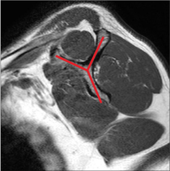
Fig. 1 Scapular Y-view. In the magnetic resonance imaging oblique sagittal view of the shoulder, the surface where the scapula and spine form a Y-shape is referred to as the scapular Y-view.
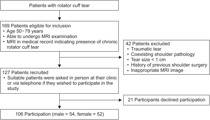
Fig. 2 Study participant recruitment flow chart. Recruitment criteria and illustration of the selection process. MRI: magnetic resonance imaging.
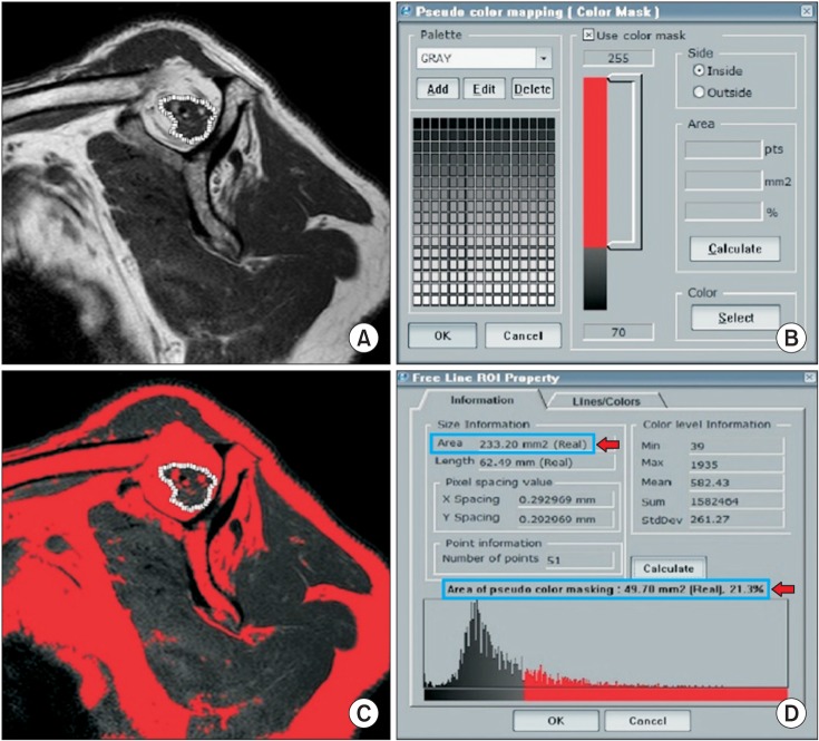
Fig. 3 Measurement of the fatty infiltration ratio in a sagittal magnetic resonance imaging image using the picture archiving and communication system computer program. (A) The area of the muscle portion in the supraspinatus muscle was established using a free line range of interest function. (B) The pseudo-color mapping function was used to establish the parts that showed a signal intensity of fat. (C) The color mapping of the fatty infiltration area was provided. (D) By calculating the color-mapped portion in the muscle, the area of muscle and fat, and their ratio could be derived.

Fig. 4 Three-slice levels in a shoulder magnetic resonance imaging oblique sagittal view. (A) The medial third slice to the scapular Y-view. (B) The scapular Y-view. (C) The lateral half slice to the scapular Y-view (in this case, the fifth slice).
Reference
-
1. Gasbarro G, Ye J, Newsome H, et al. Morphologic risk factors in predicting symptomatic structural failure of arthroscopic rotator cuff repairs: tear size, location, and atrophy matter. Arthroscopy. 2016; 32(10):1947–1952. PMID: 27129377.
Article2. Gladstone JN, Bishop JY, Lo IK, Flatow EL. Fatty infiltration and atrophy of the rotator cuff do not improve after rotator cuff repair and correlate with poor functional outcome. Am J Sports Med. 2007; 35(5):719–728. PMID: 17337727.
Article3. Goutallier D, Postel JM, Gleyze P, Leguilloux P, Van Driessche S. Influence of cuff muscle fatty degeneration on anatomic and functional outcomes after simple suture of full-thickness tears. J Shoulder Elbow Surg. 2003; 12(6):550–554. PMID: 14671517.
Article4. Le BT, Wu XL, Lam PH, Murrell GA. Factors predicting rotator cuff retears: an analysis of 1000 consecutive rotator cuff repairs. Am J Sports Med. 2014; 42(5):1134–1142. PMID: 24748610.5. Liem D, Lichtenberg S, Magosch P, Habermeyer P. Magnetic resonance imaging of arthroscopic supraspinatus tendon repair. J Bone Joint Surg Am. 2007; 89(8):1770–1776. PMID: 17671017.
Article6. McElvany MD, McGoldrick E, Gee AO, Neradilek MB, Matsen FA 3rd. Rotator cuff repair: published evidence on factors associated with repair integrity and clinical outcome. Am J Sports Med. 2015; 43(2):491–500. PMID: 24753240.7. Nardo L, Karampinos DC, Lansdown DA, et al. Quantitative assessment of fat infiltration in the rotator cuff muscles using water-fat MRI. J Magn Reson Imaging. 2014; 39(5):1178–1185. PMID: 24115490.
Article8. Goutallier D, Postel JM, Bernageau J, Lavau L, Voisin MC. Fatty muscle degeneration in cuff ruptures: pre- and postoperative evaluation by CT scan. Clin Orthop Relat Res. 1994; (304):78–83. PMID: 8020238.9. Cole BJ, McCarty LP 3rd, Kang RW, Alford W, Lewis PB, Hayden JK. Arthroscopic rotator cuff repair: prospective functional outcome and repair integrity at minimum 2-year follow-up. J Shoulder Elbow Surg. 2007; 16(5):579–585. PMID: 17629505.
Article10. DeFranco MJ, Bershadsky B, Ciccone J, Yum JK, Iannotti JP. Functional outcome of arthroscopic rotator cuff repairs: a correlation of anatomic and clinical results. J Shoulder Elbow Surg. 2007; 16(6):759–765. PMID: 18061116.
Article11. Henn RF 3rd, Kang L, Tashjian RZ, Green A. Patients' preoperative expectations predict the outcome of rotator cuff repair. J Bone Joint Surg Am. 2007; 89(9):1913–1919. PMID: 17768186.
Article12. Mellado JM, Calmet J, Olona M, et al. Surgically repaired massive rotator cuff tears: MRI of tendon integrity, muscle fatty degeneration, and muscle atrophy correlated with intraoperative and clinical findings. AJR Am J Roentgenol. 2005; 184(5):1456–1463. PMID: 15855096.
Article13. Oh JH, Kim SH, Ji HM, Jo KH, Bin SW, Gong HS. Prognostic factors affecting anatomic outcome of rotator cuff repair and correlation with functional outcome. Arthroscopy. 2009; 25(1):30–39. PMID: 19111216.
Article14. Sherman SL, Lyman S, Koulouvaris P, Willis A, Marx RG. Risk factors for readmission and revision surgery following rotator cuff repair. Clin Orthop Relat Res. 2008; 466(3):608–613. PMID: 18264848.
Article15. Spencer EE Jr, Dunn WR, Wright RW, et al. Interobserver agreement in the classification of rotator cuff tears using magnetic resonance imaging. Am J Sports Med. 2008; 36(1):99–103. PMID: 17932406.
Article16. Sugaya H, Maeda K, Matsuki K, Moriishi J. Repair integrity and functional outcome after arthroscopic double-row rotator cuff repair: a prospective outcome study. J Bone Joint Surg Am. 2007; 89(5):953–960. PMID: 17473131.17. Fuchs B, Weishaupt D, Zanetti M, Hodler J, Gerber C. Fatty degeneration of the muscles of the rotator cuff: assessment by computed tomography versus magnetic resonance imaging. J Shoulder Elbow Surg. 1999; 8(6):599–605. PMID: 10633896.
Article18. Lee E, Choi JA, Oh JH, et al. Fatty degeneration of the rotator cuff muscles on pre- and postoperative CT arthrography (CTA): is the Goutallier grading system reliable? Skeletal Radiol. 2013; 42(9):1259–1267. PMID: 23793351.
Article19. Zanetti M, Gerber C, Hodler J. Quantitative assessment of the muscles of the rotator cuff with magnetic resonance imaging. Invest Radiol. 1998; 33(3):163–170. PMID: 9525755.
Article20. Vidt ME, Santago AC 2nd, Tuohy CJ, et al. Assessments of fatty infiltration and muscle atrophy from a single magnetic resonance image slice are not predictive of 3-dimensional measurements. Arthroscopy. 2016; 32(1):128–139. PMID: 26391648.
Article21. Park JS, Park HJ, Kim SH, Oh JH. Prognostic factors affecting rotator cuff healing after arthroscopic repair in small to medium-sized tears. Am J Sports Med. 2015; 43(10):2386–2392. PMID: 26286879.
Article22. DeOrio JK, Cofield RH. Results of a second attempt at surgical repair of a failed initial rotator-cuff repair. J Bone Joint Surg Am. 1984; 66(4):563–567. PMID: 6707035.
Article23. Gerber C, Schneeberger AG, Hoppeler H, Meyer DC. Correlation of atrophy and fatty infiltration on strength and integrity of rotator cuff repairs: a study in thirteen patients. J Shoulder Elbow Surg. 2007; 16(6):691–696. PMID: 17931904.
Article24. Jost B, Pfirrmann CW, Gerber C, Switzerland Z. Clinical outcome after structural failure of rotator cuff repairs. J Bone Joint Surg Am. 2000; 82(3):304–314. PMID: 10724223.
Article25. Melis B, DeFranco MJ, Chuinard C, Walch G. Natural history of fatty infiltration and atrophy of the supraspinatus muscle in rotator cuff tears. Clin Orthop Relat Res. 2010; 468(6):1498–1505. PMID: 20094853.
Article26. Kuzel BR, Grindel S, Papandrea R, Ziegler D. Fatty infiltration and rotator cuff atrophy. J Am Acad Orthop Surg. 2013; 21(10):613–623. PMID: 24084435.
Article27. Maman E, Harris C, White L, Tomlinson G, Shashank M, Boynton E. Outcome of nonoperative treatment of symptomatic rotator cuff tears monitored by magnetic resonance imaging. J Bone Joint Surg Am. 2009; 91(8):1898–1906. PMID: 19651947.
Article28. Oh JH, Kim SH, Choi JA, Kim Y, Oh CH. Reliability of the grading system for fatty degeneration of rotator cuff muscles. Clin Orthop Relat Res. 2010; 468(6):1558–1564. PMID: 19347412.
Article29. Tingart MJ, Apreleva M, Lehtinen JT, Capell B, Palmer WE, Warner JJ. Magnetic resonance imaging in quantitative analysis of rotator cuff muscle volume. Clin Orthop Relat Res. 2003; (415):104–110. PMID: 14612636.
Article30. Thomazeau H, Rolland Y, Lucas C, Duval JM, Langlais F. Atrophy of the supraspinatus belly: assessment by MRI in 55 patients with rotator cuff pathology. Acta Orthop Scand. 1996; 67(3):264–268. PMID: 8686465.
Article31. Slabaugh MA, Friel NA, Karas V, Romeo AA, Verma NN, Cole BJ. Interobserver and intraobserver reliability of the Goutallier classification using magnetic resonance imaging: proposal of a simplified classification system to increase reliability. Am J Sports Med. 2012; 40(8):1728–1734. PMID: 22753846.
- Full Text Links
-
- Actions
-
Cited
- CITED
-
- Close
- Share
-
- Similar articles
-
- Fatty Degeneration and Atrophy of Rotator Cuffs: Comparison of Immediate Postoperative MRI with Preoperative MRI
- Partial-Thickness Tear of Supraspinatus and Infraspinatus Tendon Revisited: Based on MR Findings
- Relationship of the Sagittal Extent of Rotator Cuff Tears to the Grade of Fatty Degeneration of the Rotator Cuff Muscles
- 16-Slice MDCT Arthrography of the Shoulder: Accuracy for Detection of Glenoid Labral and Rotator Cuff Tears
- Preoperative Korean Shoulder Scoring System Correlates with Preoperative Factors of Rotator Cuff Tears
