Restor Dent Endod.
2017 Nov;42(4):324-331. 10.5395/rde.2017.42.4.324.
Smear layer removal by different chemical solutions used with or without ultrasonic activation after post preparation
- Affiliations
-
- 1Department of Restorative Dentistry, Londrina State University (UEL), Londrina, PR, Brazil.
- 2Dentistry Course, Paranaense University, Cascavel, PR, Brazil.
- 3Clinical Practice Limited to Esthetic Dentistry, Campo Grande, MS, Brazil.
- 4Clinical Practice Limited to Endodontics, Navegantes, SC, Brazil. ricardo.machado.endo@gmail.com
- 5Department of Endodontics, São Paulo State University (UNESP) School of Dentistry, Araçatuba, SP, Brazil.
- 6Department of Oral Medicine and Pediatric Dentistry, Londrina State University (UEL), Londrina, PR, Brazil.
- KMID: 2403074
- DOI: http://doi.org/10.5395/rde.2017.42.4.324
Abstract
OBJECTIVES
This study evaluated smear layer removal by different chemical solutions used with or without ultrasonic activation after post preparation.
MATERIALS AND METHODS
Forty-five extracted uniradicular human mandibular premolars with single canals were treated endodontically. The cervical and middle thirds of the fillings were then removed, and the specimens were divided into 9 groups: G1, saline solution (NaCl); G2, 2.5% sodium hypochlorite (NaOCl); G3, 2% chlorhexidine (CHX); G4, 11.5% polyacrylic acid (PAA); G5, 17% ethylenediaminetetraacetic acid (EDTA). For the groups 6, 7, 8, and 9, the same solutions used in the groups 2, 3, 4, and 5 were used, respectively, but activated with ultrasonic activation. Afterwards, the roots were analyzed by a score considering the images obtained from a scanning electron microscope.
RESULTS
EDTA achieved the best performance compared with the other solutions evaluated regardless of the irrigation method (p < 0.05).
CONCLUSIONS
Ultrasonic activation did not significantly influence smear layer removal.
Keyword
MeSH Terms
Figure
-
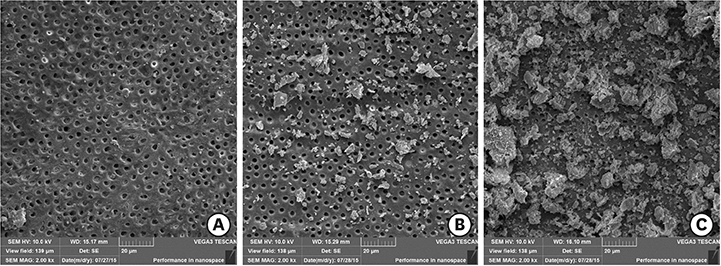
Figure 1 Representative images: (A) score 1; (B) score 2; (C) score 3.
Cited by 1 articles
-
Effect of irrigation protocols on smear layer removal, bond strength and nanoleakage of fiber posts using a self-adhesive resin cement
Rodrigo Stadler Alessi, Renata Terumi Jitumori, Bruna Fortes Bittencourt, Giovana Mongruel Gomes, João Carlos Gomes
Restor Dent Endod. 2023;48(3):e28. doi: 10.5395/rde.2023.48.e28.
Reference
-
1. Kirkevang LL, Ørstavik D, Hörsted-Bindslev P, Wenzel A. Periapical status and quality of root fillings and coronal restorations in a Danish population. Int Endod J. 2000; 33:509–515.
Article2. Hommez GM, Coppens CR, De Moor RJ. Periapical health related to the quality of coronal restorations and root fillings. Int Endod J. 2002; 35:680–689.
Article3. Mohammadi Z, Jafarzadeh H, Shalavi S, Bhandi S, Kinoshita J. Resilon: Review of a new material for obturation of the canal. J Contemp Dent Pract. 2015; 16:407–414.
Article4. Souza LC, Yadlapati M, Dorn SO, Silva R, Letra A. Analysis of radiopacity, pH and cytotoxicity of a new bioceramic material. J Appl Oral Sci. 2015; 23:383–389.
Article5. Carvalho AO, Bruzi G, Anderson RE, Maia HP, Giannini M, Magne P. Influence of adhesive core buildup designs on the resistance of endodontically treated molars restored with lithium disilicate CAD/CAM crowns. Oper Dent. 2016; 41:76–82.
Article6. Veríssimo C, Simamoto Júnior PC, Soares CJ, Noritomi PY, Santos-Filho PC. Effect of the crown, post, and remaining coronal dentin on the biomechanical behavior of endodontically treated maxillary central incisors. J Prosthet Dent. 2014; 111:234–246.
Article7. Freedman GA. Esthetic post-and-core treatment. Dent Clin North Am. 2001; 45:103–116.8. Goracci C, Ferrari M. Current perspectives on post systems: a literature review. Aust Dent J. 2011; 56:77–83.
Article9. Calixto LR, Bandéca MC, Clavijo V, Andrade MF, Vaz LG, Campos EA. Effect of resin cement system and root region on the push-out bond strength of a translucent fiber post. Oper Dent. 2012; 37:80–86.
Article10. Serafino C, Gallina G, Cumbo E, Ferrari M. Surface debris of canal walls after post space preparation in endodontically treated teeth: a scanning electron microscopic study. Oral Surg Oral Med Oral Pathol Oral Radiol Endod. 2004; 97:381–387.
Article11. Baena E, Flores A, Ceballos L. Influence of root dentin treatment on the push-out bond strength of fiber posts. Odontology. 2017; 105:170–177.
Article12. Violich DR, Chandler NP. The smear layer in endodontics - a review. Int Endod J. 2010; 43:2–15.
Article13. Kuçi A, Alaçam T, Yavaş O, Ergul-Ulger Z, Kayaoglu G. Sealer penetration into dentinal tubules in the presence or absence of smear layer: a confocal laser scanning microscopic study. J Endod. 2014; 40:1627–1631.
Article14. Scotti N, Rota R, Scansetti M, Migliaretti G, Pasqualini D, Berutti E. Fiber post adhesion to radicular dentin: The use of acid etching prior to a one-step self-etching adhesive. Quintessence Int. 2012; 43:615–623.15. Kuah HG, Lui JN, Tseng PS, Chen NN. The effect of EDTA with and without ultrasonics on removal of the smear layer. J Endod. 2009; 35:393–396.
Article16. Roy RA, Ahmad M, Crum LA. Physical mechanisms governing the hydrodynamic response of an oscillating ultrasonic file. Int Endod J. 1994; 27:197–207.
Article17. Kato AS, Cunha RS, da Silveira Bueno CE, Pelegrine RA, Fontana CE, de Martin AS. Investigation of the efficacy of passive ultrasonic irrigation versus irrigation with reciprocating activation: An environmental scanning electron microscopic study. J Endod. 2016; 42:659–663.
Article18. Torabinejad M, Cho Y, Khademi AA, Bakland LK, Shabahang S. The effect of various concentrations of sodium hypochlorite on the ability of MTAD to remove the smear layer. J Endod. 2003; 29:233–239.
Article19. Van Meerbeek B, De Munck J, Yoshida Y, Inoue S, Vargas M, Vijay P, Van Landuyt K, Lambrechts P, Vanherle G. Buonocore memorial lecture. Adhesion to enamel and dentin: current status and future challenges. Oper Dent. 2003; 28:215–235.20. Boone KJ, Murchison DF, Schindler WG, Walker WA. Post retention: the effect of sequence of post-space preparation, cementation time, and different sealers. J Endod. 2001; 27:768–771.
Article21. Lui JN, Kuah HG, Chen NN. Effect of EDTA with and without surfactants or ultrasonics on removal of smear layer. J Endod. 2007; 33:472–475.
Article22. Poggio C, Dagna A, Chiesa M, Bianchi S, Arciola CR, Visai L, Giardino L. SEM evaluation of the root canal walls after treatment with Tetraclean. Int J Artif Organs. 2010; 33:660–666.
Article23. Gu XH, Mao CY, Kern M. Effect of different irrigation on smear layer removal after post space preparation. J Endod. 2009; 35:583–586.
Article24. de Vasconcelos BC, Luna-Cruz SM, De-Deus G, de Moraes IG, Maniglia-Ferreira C, Gurgel-Filho ED. Cleaning ability of chlorhexidine gel and sodium hypochlorite associated or not with EDTA as root canal irrigants: a scanning electron microscopy study. J Appl Oral Sci. 2007; 15:387–391.
Article25. Haapasalo M, Shen Y, Qian W, Gao Y. Irrigation in endodontics. Dent Clin North Am. 2010; 54:291–312.
Article26. Lo Giudice G, Lizio A, Giudice RL, Centofanti A, Rizzo G, Runci M, Alibrandi A, Cicciù M. The effect of different cleaning protocols on post space: a SEM study. Int J Dent. 2016; 2016:1907124.
Article27. Mirseifinejad R, Tabrizizade M, Davari A, Mehravar F. Efficacy of different root canal irrigants on smear layer removal after post space preparation: a scanning electron microscopy evaluation. Iran Endod J. 2017; 12:185–190.28. Choudhary K, Nandlal B. Comparative evaluation of shear bond strength of nano-hydroxyapatite incorporated glass ionomer cement and conventional glass ionomer cement on dense synthetic hydroxyapatite disk: An in vitro study. Indian J Dent Res. 2015; 26:170–175.
Article29. Youm SH, Jung KH, Son SA, Kwon YH, Park JK. Effect of dentin pretreatment and curing mode on the microtensile bond strength of self-adhesive resin cements. J Adv Prosthodont. 2015; 7:317–322.
Article30. Mount GJ. Buonocore Memorial Lecture. Glass-ionomer cements: past, present and future. Oper Dent. 1994; 19:82–90.31. Vasiliadis L, Darling AI, Levers BG. The amount and distribution of sclerotic human root dentine. Arch Oral Biol. 1983; 28:645–649.
Article32. Vasiliadis L, Darling AI, Levers BG. The histology of sclerotic human root dentine. Arch Oral Biol. 1983; 28:693–700.
Article33. van der Sluis LW, Vogels MP, Verhaagen B, Macedo R, Wesselink PR. Study on the influence of refreshment/activation cycles and irrigants on mechanical cleaning efficiency during ultrasonic activation of the irrigant. J Endod. 2010; 36:737–740.
Article34. Sabins RA, Johnson JD, Hellstein JW. A comparison of the cleaning efficacy of short-term sonic and ultrasonic passive irrigation after hand instrumentation in molar root canals. J Endod. 2003; 29:674–678.
Article
- Full Text Links
-
- Actions
-
Cited
- CITED
-
- Close
- Share
-
- Similar articles
-
- Smear layer removal by passive ultrasonic irrigation and 2 new mechanical methods for activation of the chelating solution
- Impact of different agitation methods on smear layer cleaning of mesial canals with accentuated curvature
- Effect of irrigation protocols on smear layer removal, bond strength and nanoleakage of fiber posts using a self-adhesive resin cement
- Dentinal tubule penetration of sodium hypochlorite in root canals with and without mechanical preparation and different irrigant activation methods
- Comparison of various activation methods of root canal irrigants for soft-tissue removal
