Nat Prod Sci.
2017 Dec;23(4):270-273. 10.20307/nps.2017.23.4.270.
Quantitative Determination of Bakkenolide D in Petasites japonicus and Farfugium japonicum by HPLC/UV
- Affiliations
-
- 1Department of Integrative Plant Science, Chung-Ang University, Anseong 17546, Korea. slee@cau.ac.kr
- 2Department of Food Science and Nutrition & Kimchi Research Institute, Pusan National University, Busan 46241, Korea.
- 3Department of Food Science, Gyeongnam National University of Science and Technology, Jinju 52725, Korea. hykim@gntech.ac.kr
- KMID: 2401579
- DOI: http://doi.org/10.20307/nps.2017.23.4.270
Abstract
- A quantitative analysis of bakkenolide D in the different parts of Petasites japonicus and Farfugium japonicum was performed by HPLC. A gradient HPLC elution system with a mobile phase consisting of water:acetonitrile solution (20:80 to 0:100 for 45 min) was followed and an INNO Câ‚₈ column was used for the chromatographic separation. The injection volume, flow rate, and UV detection were 10 µL, 1 mL/min, and 290 nm, respectively. Results show that both species showed the highest amount of bakkenolide D in the roots being 107.203 and 166.103 mg/g for P. japonicas and F. japonicum, respectively. Content analysis on the different parts of both plants displayed remarkably lower values which ranged from 0.403 - 4.419 and 7.252 - 32.614 mg/g for P. japonicas and F. japonicum, respectively. The results show that the roots of both plants are rich in bakkenolide D showing a promising use in the development of nutraceuticals and industrial application of the compound.
Figure
-
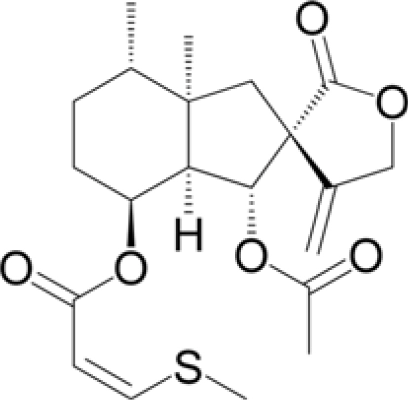
Fig. 1. Structure of bakkenolide D.

Fig. 2. HPLC chromatograms of bakkenolide D (A) and the MeOH extracts of P. japonicus (B) and F. japonicum (C).
Reference
-
References
(1). Sok D. E.., Oh S. H.., Kim Y. B.., Kang H. G.., Kim M. R.Eur. J. Nutr. 2006. 45:61–69.(2). Sugama K.., Hayashi K.., Mitsuhashi H.Phytochemistry. 1985. 24:1531–1535.(3). Okuno H.., Nakata M.., Mii M.Chromosome Sci. 2009. 12:27–33.(4). Zhao J. H.., Shen T.., Yang X.., Zhao H.., Li X.., Xie W. D.Arch. Pharm. Res. 2012. 35:1153–1158.(5). Aydin A. A.., Zerbes V.., Parlar H.., Letzel T. J.Pharm. Biomed. Anal. 2013. 75:220–229.(6). Dai D.., Pei L.., Tang L.., Chen F.., Chen X.Biomed. Chromatogr. 2013. 27:1200–1207.
Article(7). Kim, J. Y; Oh T. H.., Kim B. J.., Kim S. S.., Lee N. H.., Hyun C. G. J.Oleo Sci. 2008. 57:623–628.(8). Lee K. P.., Kang S.., Park S. J.., Choi Y. W.., Lee Y. G.., Im D. S. J.Ethnopharmacol. 2013. 148:890–894.(9). Wang S.., Jin D. Q.., Xie C.., Wang H.., Wang M.., Xu J.., Guo Y.Food Chem. 2013. 141:2075–2082.(10). Nagano H.., Moriyama Y.., Tanahashi Y.., Takahashi T.Chem. Lett. 1972. 1:13–16.(11). Hatanaka A.., Kajiwara T.., Sekiya J.., Hirata H.Agr. Biol. Chem. 1976. 40:2177–2180.(12). Niwa H.., Ishiwata H.., Yamada K. J.Nat. Prod. 1985. 48:1003–1004.(13). Tori M.., Otose K.., Fukuyama H.., Murata J.., Shiotani Y.., Takaoka S.., Nakashima K.., Sono M.., Tanaka M.Tetrahedron. 2010. 66:5235–5243.(14). Evans D. A.., Sims C. L.Tetrahedron Lett. 1973. 47:4691–4694.(15). Kim T. H.., Kim D. Y.., Jung W. J.., Nagairya R.., Son B. G.., Park Y. H.., Kang J. S.., Lee Y. J.., Im D. S.., Lee Y. G.., Choi Y. H.., Choi Y. I. J.Life Sci. 2014. 24:252–259.(16). Wang Y.., Guo M.., Zhang G.., Xue Q.Acad.J. Sec. Mil. Med. Univ. 2006. 27:1210–1213.(17). Wu T. S.., Kao M. S.., Wu P. L.., Lin F. W.., Shi L. S.., Liou M. J.., Li C. Y.Chem. Pharm. Bull. 1999. 47:375–382.(18). Wang Y. L.., Guo M.., Wang Y.Chromatographia. 2009. 70:1367–1371.(19). He J.., Wang Q.., Wang Y.., Chen R.., Zhang Y.., Guo M.Acta Pharm. Sin. B. 2013. 3:354–360.
- Full Text Links
-
- Actions
-
Cited
- CITED
-
- Close
- Share
-
- Similar articles
-
- Simultaneous Determination of Four Compounds from Cercidiphyllum japonicum Using HPLC-UV Analysis
- First Report of Leaf Rust Caused by Puccinia caricis in Farfugium japonicum in Korea
- Aqueous extract of Petasites japonicus leaves promotes osteoblast differentiation via up-regulation of Runx2 and Osterix in MC3T3-E1 cells
- Bilin and Bilinone Chlorophyll Catabolite Content in the Hamamelidaceae Autumnal Leaves
- Antimutagenic and anticarcinogenic effect of methanol extracts of Petasites japonicus Maxim leaves
