Features Causing Confusion between Basal Cell Carcinoma and Squamous Cell Carcinoma in Clinical Diagnosis
- Affiliations
-
- 1Department of Dermatology, Korea University College of Medicine, Seoul, Korea. drsshong@hanmail.net
- 2Jincheon Public Health Center, Jincheon, Korea.
- KMID: 2399756
- DOI: http://doi.org/10.5021/ad.2018.30.1.64
Abstract
- BACKGROUND
Although squamous cell carcinoma (SCC) and basal cell carcinoma (BCC) can be easily diagnosed clinically, proper diagnosis is sometimes difficult when based on clinical information alone.
OBJECTIVE
To know what causes clinical misdiagnosis between SCC and BCC, and evaluate whether dermoscopy can improve diagnostic accuracy.
METHODS
Clinical and dermoscopic photographs of inversely diagnosed cases (histologically confirmed BCC with a clinical impression of SCC or vice versa) were randomly presented to six dermatologists and the reasons for each correct or incorrect diagnosis were analyzed.
RESULTS
Among 154 cases (SCCs or BCCs), 13 cases were inversely diagnosed; 9 SCCs were clinically misdiagnosed as BCC and 4 BCCs were clinically misdiagnosed as SCC. Clinically, scales, pigmentation and rolled border were meaningful factors to discern two carcinomas. Scales without both pigmentation and rolled border was favored for SCC, but BCC favored vice versa. Ulceration, telangiectasia and translucency contributed as confusing factors for proper diagnosis. Dermoscopy improved overall diagnostic accuracy to odds ratio 2.86.
CONCLUSION
SCC has a higher tendency to be clinically misdiagnosed as BCC than vice versa. Pigmentation and rolled border are factors causing misdiagnosis of SCC as BCC and BCC may be misdiagnosed as SCC in the presence of scaling. Dermoscopy seems to improve the clinical diagnostic accuracy but has limitations for some ambiguous lesions.
MeSH Terms
Figure
-

Fig. 1 Flowchart of the study. Of a total 154 squamous cell carcinoma (SCC) and basal cell carcinoma (BCC) cases, 13 cases that were misdiagnosed clinically with the inverse impression were enrolled in this study. Lesions with a previous laser treatment did not show improved diagnostic accuracy with dermoscopy.

Fig. 2 Clinical and dermoscopic photographs of squamous cell carcinoma misdiagnosed as basal cell carcinoma. Ulcer surrounded by rolled border and arborizing vessel (A, C) in case 6, and pigmentation with telangiectasia and ulceration (B, D) in case 8 are seen.
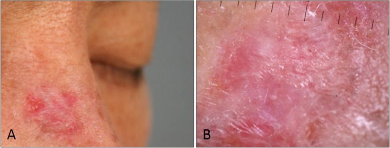
Fig. 3 Clinical and dermoscopic photographs of basal cell carcinoma (BCC) clinically misdiagnosed as squamous cell carcinoma. There are no pigmentations but some scales and vessels are present (A). Dermoscopy shows only keratin without BCC patterns such as blue-gray globules, arborizing vessels, and spoke-wheel areas, may be result of previous laser therapy (B).
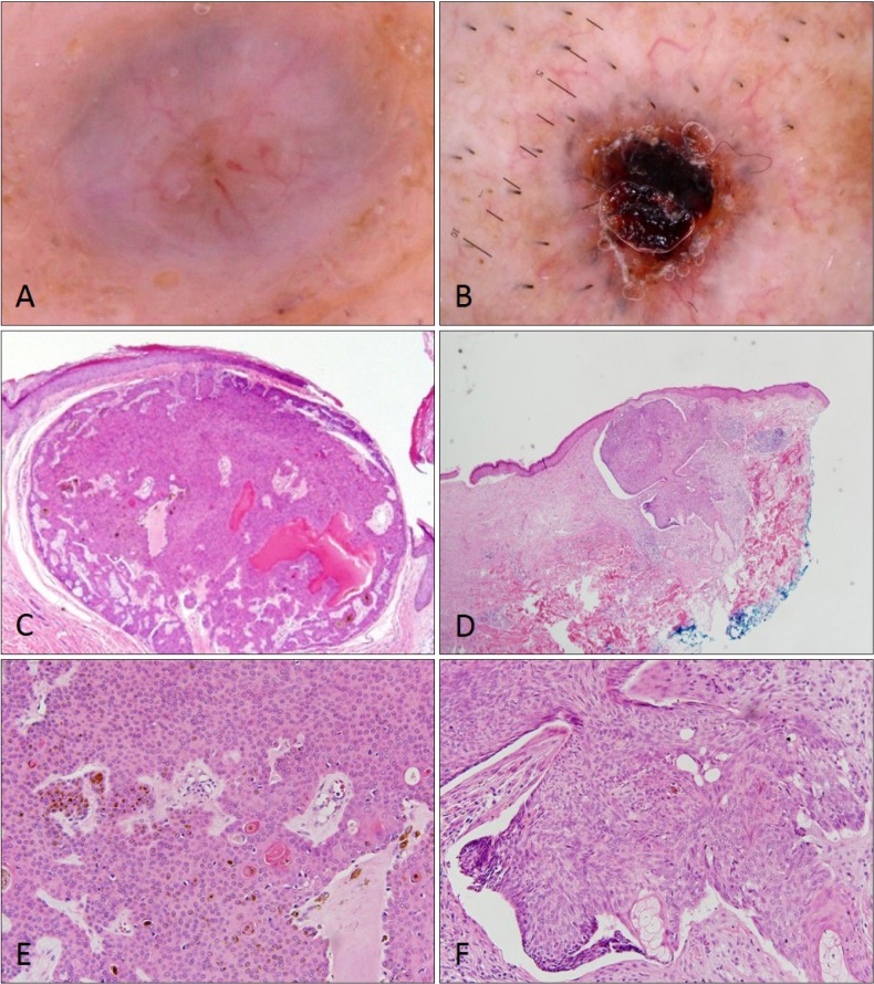
Fig. 4 Dermoscopic and histopathologic findings of basosquamous cell carcinoma. Basosquamous cell carcinoma appeared as basal cell carcinoma or squamous cell carcinoma according to histopathologic findings. Arborizing vessels with hyperkeratosis (A, C, E) and ulceration with telangiectasia (B, D, F) are seen (H&E; C, D: ×40, E, F: ×100).
Reference
-
1. Geller AC, Annas GD. Epidemiology of melanoma and nonmelanoma skin cancer. Semin Oncol Nurs. 2003; 19:2–11.
Article2. Lee KH, Lee AY, Lee CW, Park CK, Shu JI, Kim IS. Frequency trends of basal cell carcinoma, squamous cell carcinoma and melanoma in Korea, between mid-1980s and mid-1990s. Ann Dermatol. 1999; 11:70–74.
Article3. Alam M, Ratner D. Cutaneous squamous-cell carcinoma. N Engl J Med. 2001; 344:975–983. PMID: 11274625.
Article4. Rosendahl C, Cameron A, Argenziano G, Zalaudek I, Tschandl P, Kittler H. Dermoscopy of squamous cell carcinoma and keratoacanthoma. Arch Dermatol. 2012; 148:1386–1392. PMID: 22986634.
Article5. Zalaudek I, Kreusch J, Giacomel J, Ferrara G, Catricalà C, Argenziano G. How to diagnose nonpigmented skin tumors: a review of vascular structures seen with dermoscopy: part II. Nonmelanocytic skin tumors. J Am Acad Dermatol. 2010; 63:377–386. PMID: 20708470.6. Altamura D, Menzies SW, Argenziano G, Zalaudek I, Soyer HP, Sera F, et al. Dermatoscopy of basal cell carcinoma: morphologic variability of global and local features and accuracy of diagnosis. J Am Acad Dermatol. 2010; 62:67–75. PMID: 19828209.
Article7. Kim WJ, Song M, Kim HS, Ko HC, Kim MB, Kim BS. History of laser ablation in pigmented basal cell carcinoma conceals classic dermoscopic patterns. Dermatol Surg. 2014; 40:733–738. PMID: 25111344.
- Full Text Links
-
- Actions
-
Cited
- CITED
-
- Close
- Share
-
- Similar articles
-
- Comments to "A Case of Squamous Cell Carcinoma Developing in a Nevus Sebaceus of the Scalp"
- Basal Cell Carcinoma Arising in a Tattooed Eyebrow
- DNA analysis of squamous cell carcinoma and basal cell carcinoma of the skin using flow cytometry
- A case of adenoid basal cell carcinoma in uterine cervix
- Diagnosis with Reflectance Confocal Microscopy of Squamous Cell Carcinoma Mimicking Pigmented Basal Cell Carcinoma
