A Comparison of Efficacies of Aflibercept and Ranibizumab, Depending on the Angiographic Classification of Polypoidal Choroidal Vasculopathy
- Affiliations
-
- 1Department of Ophthalmology, Yeungnam University College of Medicine, Daegu, Korea. msagong@ynu.ac.kr
- KMID: 2397850
- DOI: http://doi.org/10.3341/jkos.2017.58.12.1356
Abstract
- PURPOSE
To compare the short-term efficacy of intravitreal aflibercept and ranibizumab treatment according to the subtypes of polypoidal choroidal vasculopathy (PCV) based on indocyanine green angiography (ICGA).
METHODS
Fifty-five treatment naïve patients with PCV who underwent intravitreal anti-vascular endothelial growth factor (VEGF) (ranibizumab, 26 eyes; aflibercept, 29 eyes) injection were retrospectively analyzed. Based on ICGA, subjects with feeder and draining vessels were defined as type 1 PCV (33 eyes), and subjects who did not have either feeder or draining vessels, but had branch vascular networks were defined as type 2 PCV (22 eyes). The complete polyp regression was assessed at 3 months after the initial treatment using ICGA. Changes in best-corrected visual acuity (BCVA) and optical coherence tomographic parameters were evaluated at 3 and 6 months.
RESULTS
Patients with type 1 PCV showed a higher complete polyp regression percentage (p = 0.034) and better visual improvement (p = 0.017) after three monthly injections compared to patients with Type 2 PCV. At 3 and 6 months, the BCVA was significantly improved in type 1 PCV patients, but not in type 2 PCV patients. In patients with type 1 PCV, the aflibercept-treated group showed a better response in anatomical outcomes (p = 0.020), and complete polyp regression percentage (p = 0.027; dry macula) than the ranibizumab-treated group, and only the aflibercept-treated group showed a significant improvement of BCVA at 3 and 6 months. In patients with type 2 PCV, there were no significant differences in visual and anatomical outcome between the anti-VEGF agents.
CONCLUSIONS
Type 1 PCV showed better visual improvement with a higher percentage of polyp regression than type 2 PCV. Anatomical changes were greater in patients treated with aflibercept than with ranibizumab, particularly in patients with type 1 PCV. These results suggest that a consideration of angiographic features is important in establishing a treatment strategy for patients with PCV.
Keyword
MeSH Terms
Figure
-

Figure 1 Morphological classification of polypoidal choroidal vasculopathy (PCV) based on indocyanine green angiography (ICGA). ICGA of a patient with type 1 polypoidal choroidal neovascularization (CNV), a feeder vessel (white arrowheads) is visible at 20s (A). At 59s (B), a drainage vessel is observed (white arrows). In the subtraction (A from B) image (C), the feeder vessel was not showed but the drainage vessel (white arrows) was visible. ICGA of a patient with type 2 polypoidal CNV, the small weak branch vascular network (BVN) (black arrow) and choroidal vessels are enhanced at 23s (D). At 58s (E), a polyp and small BVN (black arrow) are more prominent. In the subtraction (D from E) image (F), the small BVN (black arrow) was still visible, which indicates slowing of the vessel filling.
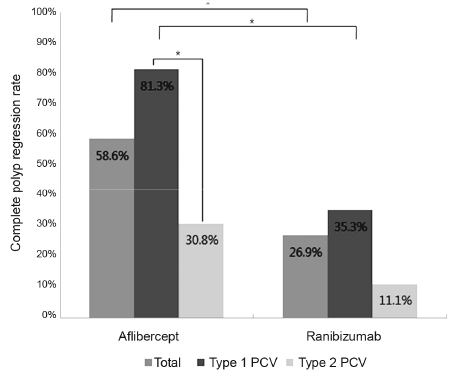
Figure 2 Comparison of the complete polyp regression between the aflibercept-and ranibizumab-treated groups. The aflibercept-treated group showed a better polyp regression than the ranibizumab-treated group in patients with type 1 polypoidal choroidal vasculopathy (PCV) (p = 0.020), while patients with type 2 PCV showed no significant difference between the two groups (p = 0.436). Patients with type 1 PCV showed a significantly higher regression percentage than patients with type 2 PCV in the aflibercept-treated group (p = 0.020), while the ranibizumab-treated group showed no significant difference between PCV types (p = 0.370). *p < 0.05, Fisher's exact test.
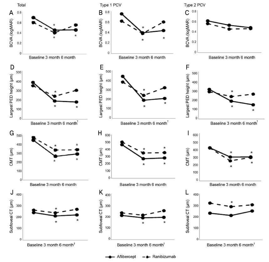
Figure 3 Outcomes of the 6-month treatment effects and comparisons between anti-vascular endothelial growth factor (anti-VEGF) agents. The aflibercept-treated group showed a better response than the ranibizumab-treated group (A, D, G, J). The anatomical changes were greater in patients treated with aflibercept than ranibizumab, particularly in patients with type 1 polypoidal choroidal vasculopathy (PCV) (B, E, H, K). In patients with type 2 PCV (C, F, I, L), there was no significant visual and anatomical difference between the anti-VEGF agents. BCVA = best-corrected visual acuity; PED = pigment epithelial detachment; CMT = central macular thickness; CT = choroidal thickness. *Parameters significantly changed from baseline (p < 0.05, Mann-Whitney U-test); †Changes in treatment effects that were significantly different between the anti-VEGF agents (p < 0.05, Mann-Whitney U test).
Reference
-
1. Yannuzzi LA, Sorenson J, Spaide RF, Lipson B. Idiopathic polypoidal choroidal vasculopathy (IPCV). Retina. 1990; 10:1–8.2. Spaide RF, Yannuzzi LA, Slakter JS, et al. Indocyanine green videoangiography of idiopathic polypoidal choroidal vasculopathy. Retina. 1995; 15:100–110.3. Cho HJ, Kim JW, Lee DW, et al. Intravitreal bevacizumab and ranibizumab injections for patients with polypoidal choroidal vasculopathy. Eye (Lond). 2012; 26:426–433.4. Koh A, Lee WK, Chen LJ, et al. EVEREST study: efficacy and safety of verteporfin photodynamic therapy in combination with ranibizumab or alone versus ranibizumab monotherapy in patients with symptomatic macular polypoidal choroidal vasculopathy. Retina. 2012; 32:1453–1464.5. Koh AH, Chen LJ, Chen SJ, et al. Polypoidal choroidal vasculopathy: evidence-based guidelines for clinical diagnosis and treatment. Retina. 2013; 33:686–716.6. Stewart MW. Aflibercept (VEGF Trap-eye): the newest anti-VEGF drug. Br J Ophthalmol. 2012; 96:1157–1158.7. Tan CS, Lim TH, Hariprasad SM. Current management of polypoidal choroidal vasculopathy. Ophthalmic Surg Lasers Imaging Retina. 2015; 46:786–791.8. Dhalla MS, Shah GK, Blinder KJ, et al. Combined photodynamic therapy with verteporfin and intravitreal bevacizumab for choroidal neovascularization in age-related macular degeneration. Retina. 2006; 26:988–993.9. Wong CW, Yanagi Y, Lee WK, et al. Age-related macular degeneration and polypoidal choroidal vasculopathy in Asians. Prog Retin Eye Res. 2016; 53:107–139.10. Inoue M, Yamane S, Taoka R, et al. Aflibercept for polypoidal choroidal vasculopathy: as needed versus fixed interval dosing. Retina. 2016; 36:1527–1534.11. Introini U, Casalino G, Triolo G, et al. Stereotactic radiotherapy for polypoidal choroidal vasculopathy: a pilot study. Ophthalmologica. 2015; 233:82–88.12. Oishi A, Tsujikawa A, Yamashiro K, et al. One-year result of aflibercept treatment on age-related macular degeneration and predictive factors for visual outcome. Am J Ophthalmol. 2015; 159:853–860.e1.13. Yamamoto A, Okada AA, Kano M, et al. One-year results of intravitreal aflibercept for polypoidal choroidal vasculopathy. Ophthalmology. 2015; 122:1866–1872.14. Takayama K, Kaneko H, Kataoka K, et al. Comparison between 1-year outcomes of aflibercept with and without photodynamic therapy for polypoidal choroidal vasculopathy: Retrospective observation study. PloS one. 2017; 12:e0176100.15. Kawamura A, Yuzawa M, Mori R, et al. Indocyanine green angiographic and optical coherence tomographic findings support classification of polypoidal choroidal vasculopathy into two types. Acta ophthalmol. 2013; 91:e474–e481.16. Yuzawa M, Mori R, Kawamura A. The origins of polypoidal choroidal vasculopathy. Br J Ophthalmol. 2005; 89:602–607.17. Tsujikawa A, Sasahara M, Otani A, et al. Pigment epithelial detachment in polypoidal choroidal vasculopathy. Am J Ophthalmol. 2007; 143:102–111.18. Tong JP, Chan WM, Liu DT, et al. Aqueous humor levels of vascular endothelial growth factor and pigment epithelium–derived factor in polypoidal choroidal vasculopathy and choroidal neovascularization. Am J Ophthalmol. 2006; 141:456–462.19. Tan CS, Ngo WK, Lim LW, Lim TH. A novel classification of the vascular patterns of polypoidal choroidal vasculopathy and its relation to clinical outcomes. Br J Ophthalmol. 2014; 98:1528–1533.20. Sasahara M, Tsujikawa A, Musashi K, et al. Polypoidal choroidal vasculopathy with choroidal vascular hyperpermeability. Am J Ophthalmol. 2006; 142:601–607.21. Matsumiya W, Honda S, Otsuka K, et al. Comparison of the effectiveness and prognostic factors of intravitreal ranibizumab between typical neovascular age-related macular degeneration and polypoidal choroidal vasculopathy over 24 months of follow-up. Ophthalmologica. 2015; 234:33–39.22. Koizumi H, Yamagishi T, Yamazaki T, Kinoshita S. Relationship between clinical characteristics of polypoidal choroidal vasculopathy and choroidal vascular hyperpermeability. Am J Ophthalmol. 2013; 155:305–313.e1.23. Kim JH, Lee TG, Chang YS, et al. Short-term choroidal thickness changes in patients treated with either ranibizumab or aflibercept: a comparative study. Br J Ophthalmol. 2016; 100:1634–1639.24. Jeong S, Sagong M. Short-term efficacy of intravitreal aflibercept depending on angiographic classification of polypoidal choroidal vasculopathy. Br J Ophthalmol. 2017; 101:758–763.25. Lee D, Jeong S, Moon J, et al. Analysis of efficacy of intravitreal aflibercept according to subfoveal choroidal thickness in polypoidal choroidal vasculopathy. J Korean Ophthalmol Soc. 2016; 57:1577–1585.26. Coscas G, Lupidi M, Coscas F, et al. Toward a specific classification of polypoidal choroidal vasculopathy: idiopathic disease or subtype of age-related macular degeneration. Invest Ophthalmol Vis Sci. 2015; 56:3187–3195.27. Chung SE, Kang SW, Lee JH, Kim YT. Choroidal thickness in polypoidal choroidal vasculopathy and exudative age-related macular degeneration. Ophthalmology. 2011; 118:840–845.28. Klettner A, Recber M, Roider J. Comparison of the efficacy of aflibercept, ranibizumab, and bevacizumab in an RPE/choroid organ culture. Graefes Arch Clin Exp Ophthalmol. 2014; 252:1593–1598.29. Lommatzsch A, Heimes B, Gutfleisch M, et al. Serous pigment epithelial detachment in age-related macular degeneration: comparison of different treatments. Eye (Lond). 2009; 23:2163–2168.30. Browning DJ, Kaiser PK, Rosenfeld PJ, Stewart MW. Aflibercept for age-related macular degeneration: a game-changer or quiet addition? Am J Ophthalmol. 2012; 154:222–226.31. Nomura Y, Kaneko M, Miyata K, et al. Bevacizumab and aflibercept activate platelets via FcγRIIa. Invest Ophthalmol Vis Sci. 2015; 56:8075–8082.32. Julien S, Biesemeier A, Taubitz T, Schraermeyer U. Different effects of intravitreally injected ranibizumab and aflibercept on retinal and choroidal tissues of monkey eyes. Br J Ophthalmol. 2014; 98:813–825.
- Full Text Links
-
- Actions
-
Cited
- CITED
-
- Close
- Share
-
- Similar articles
-
- Initial Factors Associated with Resistance to Intravitreal Aflibercept Injection in Polypoidal Choroidal Vasculopathy
- Comparison of Choroidal Thickness Change between Ranibizumab and Aflibercept in Age-related Macular Degeneration: Six Month Results
- Long-term Treatment Outcome of Intravitreal Aflibercept Monotherapy for Polypoidal Choroidal Vasculopathy
- Aflibercept Treatment for Neovascular Age-related Macular Degeneration and Polypoidal Choroidal Vasculopathy Refractory to Anti-vascular Endothelial Growth Factor
- Clinical Outcomes of Switching to Brolucizumab in Refractory Polypoidal Choroidal Vasculopathy Treated with Aflibercept
