Parotid mandibular bone defect: A case report emphasizing imaging features in plain radiographs and magnetic resonance imaging
- Affiliations
-
- 1Department of Oral and Maxillofacial Radiology, School of Dentistry, Okayama University, Okayama, Japan.
- 2Department of Stomatology, School of Dentistry, São Paulo University, São Paulo, SP, Brazil. dra.lucimunhoz@usp.br
- KMID: 2397842
- DOI: http://doi.org/10.5624/isd.2017.47.4.269
Abstract
- Mandibular bone depression, also known as Stafne bone cavity, is defined as a bone depression filled mainly with salivary gland tissue. Parotid gland bone defects are infrequently observed. We report the case of a 52-year-old male patient who underwent radiographic examinations due to temporomandibular joint dysfunction, and a radiolucent area was detected in the mandibular ramus, with a provisional diagnosis of traumatic bone cyst or parotid mandibular bone defect. The patient was then referred for magnetic resonance imaging, which demonstrated a hyperintense area eroding the mandibular ramus, which corresponded to glandular tissue. Although the defect was a benign lesion, radiolucencies in the mandibular ramus lead to concerns among professionals, because their radiographic features can resemble various intrabony neoplastic lesions, such as giant cell tumors or benign tumors of the parotid gland.
MeSH Terms
Figure
-
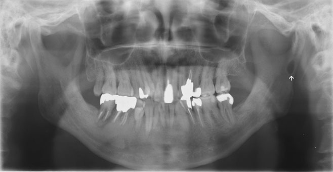
Fig. 1 Panoramic radiograph shows the ovoid radiolucent area in the left mandibular ramus (arrow).
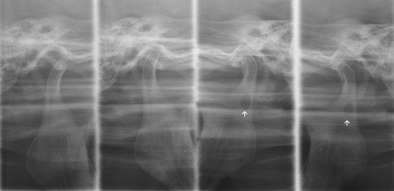
Fig. 2 Four-part panoramic radiograph of the temporomandibular joint. Views of the temporomandibular joint with the mouth open and closed demonstrate the oval-shaped radiolucency in the left side of the mandibular ramus.
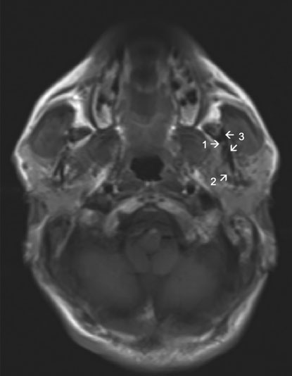
Fig. 3 An axial scout magnetic resonance image demonstrating a tiny hyperintense area in the left mandibular ramus, facing the lingual side (arrow number 1). Note the parotid glandular tissue (arrow number 2), which is also hyperintense. Arrow number 3 indicates the mandibular ramus.

Fig. 4 A proton-weighted magnetic resonance image reveals a hyperintense signal area on the left mandibular ramus (arrow).
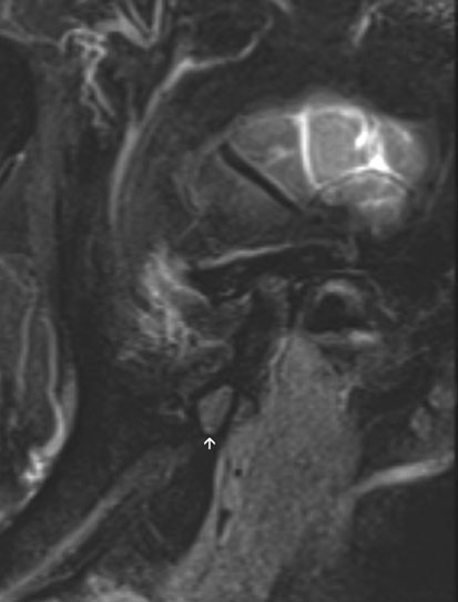
Fig. 5 A T2-weighted magnetic resonance image shows a hyperintense signal area on the left mandibular ramus, corresponding to the defect in the mandibular ramus (arrow).
Reference
-
1. Stafne EC. Bone cavities situated near the angle of the mandible. J Am Dent Assoc. 1942; 29:1969–1972.
Article2. Kaya M, Ugur KS, Dagli E, Kurtaran H, Gunduz M. Stafne bone cavity containing ectopic parotid gland. Braz J Otorhinolaryngol. (in press).
Article3. Quesada-Gómez C, Valmaseda-Castellón E, Berini-Aytés L, Gay-Escoda C. Stafne bone cavity: a retrospective study of 11 cases. Med Oral Patol Oral Cir Bucal. 2006; 11:E277–E280.4. Taysi M, Ozden C, Cankaya B, Olgac V, Yıldırım S. Stafne bone defect in the anterior mandible. Dentomaxillofac Radiol. 2014; 43:20140075.
Article5. Minowa K, Inoue N, Sawamura T, Matsuda A, Totsuka Y, Nakamura M. Evaluation of static bone cavities with CT and MRI. Dentomaxillofac Radiol. 2003; 32:2–7.
Article6. Philipsen HP, Takata T, Reichart PA, Sato S, Suei Y. Lingual and buccal mandibular bone depressions: a review based on 583 cases from a world-wide literature survey, including 69 new cases from Japan. Dentomaxillofac Radiol. 2002; 31:281–290.
Article7. Lee JI, Kang SJ, Jeon SP, Sun H. Stafne Bone cavity of the mandible. Arch Craniofac Surg. 2016; 17:162–164.
Article8. Campos PS, Panella J, Crusoé-Rebello IM, Azevedo RA, Pena N, Cunha T. Mandibular ramus-related Stafne’s bone cavity. Dentomaxillofac Radiol. 2004; 33:63–66.
Article9. Hansson LG. Development of a lingual mandibular bone cavity in an 11-year-old boy. Oral Surg Oral Med Oral Pathol. 1980; 49:376–378.
Article10. Bornstein MM, Wiest R, Balsiger R, Reichart PA. Anterior Stafne’s bone cavity mimicking a periapical lesion of endodontic origin: report of two cases. J Endod. 2009; 35:1598–1602.
Article11. Sisman Y, Etöz OA, Mavili E, Sahman H, Tarim Ertas E. Anterior Stafne bone defect mimicking a residual cyst: a case report. Dentomaxillofac Radiol. 2010; 39:124–126.
Article12. Assaf AT, Solaty M, Zrnc TA, Fuhrmann AW, Scheuer H, Heiland M, et al. Prevalence of Stafne’s bone cavity - retrospective analysis of 14,005 panoramic views. In Vivo. 2014; 28:1159–1164.13. Krafft T, Eggert J, Karl M. A Stafne bone defect in the anterior mandible - a diagnostic dilemma. Quintessence Int. 2010; 41:391–393.14. Friedrich RE, Zustin J, Scheuer HA, Assaf AT, Gröbe A. An unilateral basal bone defect of the mandible occupied by fatty tissue: Stafne’s cavity. In Vivo. 2012; 26:1045–1048.15. Etöz M, Etöz OA, Sahman H, Sekerci AE, Polat HB. An unusual case of multilocular Stafne bone cavity. Dentomaxillofac Radiol. 2012; 41:75–78.
Article16. Mauprivez C, Sahli Amor M, Khonsari RH. Magnetic resonance sialography of bilateral Stafne bone cavities. J Oral Maxillofac Surg. 2015; 73:934.e1–934.e7.
Article17. Chen CA, Ahn Y, Odell S, Mupparapu M, Graham DM. Developmental salivary gland depression in the ascending mandibular ramus: a cone-beam computed tomography study. Imaging Sci Dent. 2016; 46:223–227.
Article18. Sisman Y, Miloglu O, Sekerci AE, Yilmaz AB, Demirtas O, Tokmak TT. Radiographic evaluation on prevalence of Stafne bone defect: a study from two centres in Turkey. Dentomaxillofac Radiol. 2012; 41:152–158.
Article19. Wolf J. Bone defects in mandibular ramus resembling developmental bone cavity (Stafne). Proc Finn Dent Soc. 1985; 81:215–221.20. Barker GR. A radiolucency of the ascending ramus of the mandible associated with invested parotid salivary gland material and analogous with a Stafne bone cavity. Br J Oral Maxillofac Surg. 1988; 26:81–84.
Article21. Shields ED. Technical note: Stafne static mandibular bone defect-further expression on the buccal aspect of the ramus. Am J Phys Anthropol. 2000; 111:425–427.
Article22. Kahn JL, Bourjat P, Barrière P. Ectopic parotid gland in the mandible and masseter muscle. Rev Stomatol Chir Maxillofac. 2002; 103:195–197.23. Lee KH, Thiruchelvam JK, McDermott P. An unusual presentation of Stafne bone cyst. J Maxillofac Oral Surg. 2015; 14:841–844.
Article24. Langlais RP, Glass BJ, Bricker SL, Miles DA. Medial sigmoid depression: a panoramic pseudoforamen in the upper ramus. Oral Surg Oral Med Oral Pathol. 1983; 55:635–638.
Article25. Deyhimi P, Darisavi S, Khalesi S. Stafne bone cavity with ectopic salivary gland tissue in the anterior of mandible. Dent Res J (Isfahan). 2016; 13:454–457.
- Full Text Links
-
- Actions
-
Cited
- CITED
-
- Close
- Share
-
- Similar articles
-
- Bilateral Warthin tumor of the parotid gland: A case report
- Peritumoral Bone Marrow Edema in Benign Giant Cell Tumor
- Pediatric Cartilaginous Tibia Eminence Fracture Overlooked on Plain Radiograph: A Report of Two Cases
- Multimodality Imaging of Pediatric Parotid Gland Lesions
- Dermoid Cyst of the Parotid Gland: A Case Report
