Restor Dent Endod.
2015 Aug;40(3):216-222. 10.5395/rde.2015.40.3.216.
Evaluation of the effects of two novel irrigants on intraradicular dentine erosion, debris and smear layer removal
- Affiliations
-
- 1Department of Endodontics, Faculty of Dentistry, Hacettepe University, Ankara, Turkey. selenkkkaya@yahoo.com
- 2RAK College of Dental Science, RAK Medical and Health Science University, Ras Al Khaimah, UAE.
- 3Chemistry Department, Faculty of Science, Hacettepe University, Ankara, Turkey.
- 4Department of Endodontics, Sharjah University Dental College, Sharjah, UAE.
- KMID: 2396465
- DOI: http://doi.org/10.5395/rde.2015.40.3.216
Abstract
OBJECTIVES
To evaluate the effects of copolymer of acrylic acid and maleic acid (Poly[AA-co-MA]) and calcium hypochlorite (Ca(OCl)2) on root canal dentin using scanning electron microscope (SEM).
MATERIALS AND METHODS
Twenty-four single-rooted teeth were instrumented and the apical and coronal thirds of each root were removed, leaving the 5 mm middle thirds, which were then separated into two pieces longitudinally. The specimens were randomly divided into six groups and subjected to each irrigant for 5 min as follows: G1, Ca(OCl)2; G2, Poly(AA-co-MA); G3, Ca(OCl)2 + Poly(AA-co-MA); G4, sodium hypochlorite (NaOCl); G5, ethylenediaminetetraacetic acid (EDTA); G6, NaOCl+EDTA. The specimens were prepared for SEM evaluation. Smear layer, debris and erosion scores were recorded by two blinded examiners. One image from G3 was analyzed with energy dispersive spectroscopy (EDS) on suspicion of precipitate formation. Data were analyzed using the Kruskal-Wallis and Dunn tests.
RESULTS
G1 and G4 showed the presence of debris and smear layer and they were statistically different from G2, G3, G5 and G6 where debris and smear layer were totally removed (p < 0.05). In G1 and G4, erosion evaluation could not be done because of debris and smear layer. G2, G3 and G5 showed no erosion, and there was no significant difference between them. G6 showed severe erosion and was statistically different from G2, G3 and G5 (p < 0.05). EDS microanalysis showed the presence of Na, P, and Ca elements on the surface.
CONCLUSIONS
Poly(AA-co-MA) is effective in removing the smear layer and debris without causing erosion either alone or with Ca(OCl)2.
Keyword
MeSH Terms
Figure
-
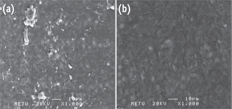
Figure 1 Photomicrographs showing the presence of debris and smear layer. (a) G1; (b) G4 (Bar = 10 µm; original magnification, ×1,000).
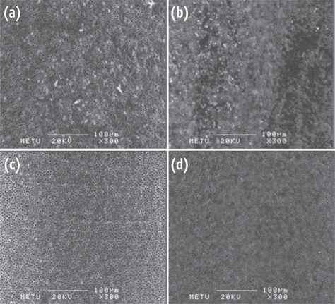
Figure 2 Photomicrographs showing the smear-free surfaces. (a) G2; (b) G3; (c) G5; (d) G6 (Bar = 100 µm; original magnification, ×300).
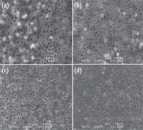
Figure 3 Representative photomicrographs for erosion evaluation. No erosion is present in (a) G2; (b) G3; (c) G5. Moderate erosion can be seen in (d) G6 (Bar = 10 µm; original magnification, ×1,000).

Figure 4 Representative photomicrographs of G6 showing severe erosion. (a) Bar = 10 µm, original magnification, ×1,000; (b) Bar = 10 µm, original magnification, ×3,000.
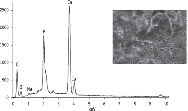
Figure 5 EDS microanalysis of a sample from G3.
Reference
-
1. Schilder H. Cleaning and shaping the root canal. Dent Clin North Am. 1974; 18:269–296.2. McComb D, Smith DC. A preliminary scanning electron microscopic study of root canals after endodontic procedures. J Endod. 1975; 1:238–242.
Article3. Peters OA, Schönenberger K, Laib A. Effects of four Ni-Ti preparation techniques on root canal geometry assessed by micro computed tomography. Int Endod J. 2001; 34:221–230.
Article4. Goldman LB, Goldman M, Kronman JH, Lin PS. The efficacy of several irrigating solutions for endodontics: a scanning electron microscopic study. Oral Surg Oral Med Oral Pathol. 1981; 52:197–204.
Article5. McComb D, Smith DC, Beagrie GS. The results of in vivo endodontic chemomechanical instrumentation-a scanning electron microscopic study. J Br Endod Soc. 1976; 9:11–18.
Article6. Gwinnett AJ. Smear layer: morphological considerations. Oper Dent Suppl. 1984; 3:2–12.7. Torabinejad M, Handysides R, Khademi AA, Bakland LK. Clinical implications of the smear layer in endodontics: a review. Oral Surg Oral Med Oral Pathol Oral Radiol Endod. 2002; 94:658–666.
Article8. Zehnder M. Root canal irrigants. J Endod. 2006; 32:389–398.
Article9. Becking AG. Complications in the use of sodium hypochlorite during endodontic treatment. Report of three cases. Oral Surg Oral Med Oral Pathol. 1991; 71:346–348.
Article10. Cobankara FK, Ozkan HB, Terlemez A. Comparison of organic tissue dissolution capacities of sodium hypochlorite and chlorine dioxide. J Endod. 2010; 36:272–274.
Article11. Dutta A, Saunders WP. Comparative evaluation of calcium hypochlorite and sodium hypochlorite on soft-tissue dissolution. J Endod. 2012; 38:1395–1398.
Article12. Mader CL, Baumgartner JC, Peters DD. Scanning electron microscopic investigation of the smeared layer on root canal walls. J Endod. 1984; 10:477–483.
Article13. Sceiza MF, Daniel RL, Santos EM, Jaeger MM. Cytotoxic effects of 10% citric acid and EDTA-T used as root canal irrigants: an in vitro analysis. J Endod. 2001; 27:741–743.14. Segura-Egea JJ, Jiménez-Rubio A, Rios-Santos JV, Velasco-Ortega E, Calvo-Gutierrez JR. In vitro inhibitory effect of EGTA on macrophage adhesion: endodontic implications. J Endod. 2003; 29:211–213.15. Baumgartner JC, Mader CL. A scanning electron microscopic evaluation of four root canal irrigation regimens. J Endod. 1987; 13:147–157.
Article16. Calt S, Serper A. Smear layer removal by EGTA. J Endod. 2000; 26:459–461.
Article17. Popescu I, Suflet DM, Pelin IM, Chitanu GC. Biomedical applications of maleic anhydride copolymers. Revue Roumaine de Chimie. 2011; 56:173–188.18. Paqué F, Barbakow F, Peters OA. Root canal preparation with Endo-Eze AET: changes in root canal shape assessed by micro-computed tomography. Int Endod J. 2005; 38:456–464.
Article19. Hülsmann M, Rümmelin C, Schäfers F. Root canal cleanliness after preparation with different endodontic handpieces and hand instruments: a comparative SEM investigation. J Endod. 1997; 23:301–306.
Article20. Torabinejad M, Khademi AA, Babagoli J, Cho Y, Johnson WB, Bozhilov K, Kim J, Shabahang S. A new solution for the removal of the smear layer. J Endod. 2003; 29:170–175.
Article21. Goldman M, Goldman LB, Cavaleri R, Bogis J, Lin PS. The efficacy of several endodontic irrigating solutions: a scanning electron microscopic study: Part 2. J Endod. 1982; 8:487–492.
Article22. Yamada RS, Armas A, Goldman M, Lin PS. A scanning electron microscopic comparison of a high volume final flush with several irrigating solutions: Part 3. J Endod. 1983; 9:137–142.
Article23. Niu W, Yoshioka T, Kobayashi C, Suda H. A scanning electron microscopic study of dentinal erosion by final irrigation with EDTA and NaOCl solutions. Int Endod J. 2002; 35:934–939.
Article24. Cruz-Filho AM, Sousa-Neto MD, Saquy PC, Pécora JD. Evaluation of the effect of EDTAC, CDTA, and EGTA on radicular dentin microhardness. J Endod. 2001; 27:183–184.
Article25. Seidberg BH, Schilder H. An evaluation of EDTA in endodontics. Oral Surg Oral Med Oral Pathol. 1974; 37:609–620.
Article26. De-Deus G, Reis C, Paciornik S. Critical appraisal of published smear layer-removal studies: methodological issues. Oral Surg Oral Med Oral Pathol Oral Radiol Endod. 2011; 112:531–543.
Article27. Peters OA, Peters CI, Schönenberger K, Barbakow F. ProTaper rotary root canal preparation: effects of canal anatomy on final shape analysed by micro CT. Int Endod J. 2003; 36:86–92.
Article28. Taha NA, Ozawa T, Messer HH. Comparison of three techniques for preparing oval-shaped root canals. J Endod. 2010; 36:532–535.
Article29. Do Prado M, Simão RA, Gomes BP. Evaluation of different irrigation protocols concerning the formation of chemical smear layer. Microsc Res Tech. 2013; 76:196–200.
Article30. Luessen HL, Borchard G, De Boer AG, Junginger HE, Kolter K, Schehlmann V, Sanner A. Use of (meth) acrylic acid/maleic acid copolymers for improving mucosal permeability. USA. US patent 6004575 A. 1999. 12. 21.
- Full Text Links
-
- Actions
-
Cited
- CITED
-
- Close
- Share
-
- Similar articles
-
- Time-dependent effects of EDTA application on removal of smear layer in the root canal system
- Debris removal efficiency depend on different ultrasonic irrigation protocols
- Removal patterns of smear layer according to application temperature and time of EDTA
- Effect of QMix irrigant in removal of smear layer in root canal system: a systematic review of in vitro studies
- The efficacy of ultrasonic irrigation technique on debris removal during root canal treatment
