Soft tissue consideration in oral rehabilitation using implant in a patient after oral tumor resection
- Affiliations
-
- 1Department of Prosthodontics and Research Institute of Oral Science, College of Dentistry, Gangneung-Wonju National University, Gangneung, Republic of Korea. lila@gwnu.ac.kr
- KMID: 2393919
- DOI: http://doi.org/10.4047/jkap.2017.55.4.458
Abstract
- After the resection of oral tumor, defected maxillofacial structure caused functional difficulties including phonetics, mastication and esthetic aspects. In this cases, implant retained prosthesis can contribute to the functional enhancement. Regardless of the success rate in grafted bone, however, the soft tissue usually had a shape which was susceptible to inflammation. Moreover, infected graft bone presented rapid destruction. For success of the prosthetic treatment, adequate soft tissue treatment and frequent recall check are the essential factors to the successful implant prognosis.
MeSH Terms
Figure
-
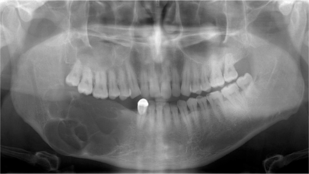
Fig. 1. Pre-operative panoramic radiograph.

Fig. 2. Post-operative panoramic radiograph.

Fig. 3. Implant installation state.

Fig. 4. Restorative procedures. (A) Impression taking, (B) Interocclusal relationship registration, (C) Wax-denture try-in, (D) Framework try-in.

Fig. 5. Implant failure state.

Fig. 6. Pre-operative panoramic radiograph.

Fig. 7. Post-operative panoramic radiograph.

Fig. 8. Panoramic radiograph of Iliac bone graft.

Fig. 9. Implant installation.

Fig. 10. Restorative procedures. (A) Impression taking, (B) Hader bar attachment, (C) Interocclusal relationship registration, (D) Definitive prosthesis.

Fig. 11. T-scan data. (A) After 2 month, (B) After 5 month, (C) After 8 month.

Fig. 12. Pre-operative panoramic radiograph.

Fig. 13. Post-operative panoramic radiograph.

Fig. 14. Implant installation state.

Fig. 15. Vestibuloplasty. (A) Pre-operation, (B) Post-operation, (C) Healing state.

Fig. 16. Restorative procedures. (A) Implant level impression, (B) Evaluation of attachment space.

Fig. 17. Customized abutment fabrication. (A) Implant installation axis, (B) Fabricated customized abutment, (C) Hader bar attachment.

Fig. 18. Registration of palatal morphology shaped by tongue movement during speech. (A) Large alveolar bone defect, (B) Record of palatal morphology using tissue conditioner.

Fig. 19. Definitive prosthesis.
Reference
-
1.Beumer J. Maxillofacial rehabilitation: prosthodontic and surgical considerations. St. Louis: Ishiyaku EuroAmerica, Co.;1996.2.Garrett N., Roumanas ED., Blackwell KE., Freymiller E., Abemayor E., Wong WK., Gerratt B., Berke G., Beumer J 3rd., Kapur KK. Efficacy of conventional and implant-supported mandibular resection prostheses: study overview and treatment outcomes. J Prosthet Dent. 2006. 96:13–24.
Article3.Tang JA., Rieger JM., Wolfaardt JF. A review of functional outcomes related to prosthetic treatment after maxillary and mandibular reconstruction in patients with head and neck cancer. Int J Prosthodont. 2008. 21:337–54.4.Bodard AG., Bémer J., Gourmet R., Lucas R., Coroller J., Salino S., Breton P. Dental implants and free fibula flap: 23 patients. Rev Stomatol Chir Maxillofac. 2011. 112:e1–4.5.Bodard AG., Salino S., Desoutter A., Deneuve S. Assessment of functional improvement with implant-supported prosthetic rehabilitation after mandibular reconstruction with a microvascular free fibula flap: A study of 25 patients. J Prosthet Dent. 2015. 113:140–5.
Article6.Parbo N., Murra NT., Andersen K., Buhl J., Kiil B., Nørholt SE. Outcome of partial mandibular reconstruction with fibula grafts and implant-supported prostheses. Int J Oral Maxillofac Surg. 2013. 42:1403–8.
Article7.Chiapasco M., Colletti G., Romeo E., Zaniboni M., Brusati R. Longterm results of mandibular reconstruction with autogenous bone grafts and oral implants after tumor resection. Clin Oral Implants Res. 2008. 19:1074–80.
Article8.Zou D., Huang W., Wang F., Wang S., Zhang Z., Zhang C., Kaigler D., Wu Y. Autologous ilium grafts: Long-term results on immediate or staged functional rehabilitation of mandibular segmental de-fects using dental implants after tumor resection. Clin Implant Dent Relat Res. 2015. 17:779–89.
Article9.Raoul G., Ruhin B., Briki S., Lauwers L., Haurou Patou G., Capet JP., Maes JM., Ferri J. Microsurgical reconstruction of the jaw with fibular grafts and implants. J Craniofac Surg. 2009. 20:2105–17.
Article10.Smith GI., O'Brien CJ., Choy ET., Andruchow JL., Gao K. Clinical outcome and technical aspects of 263 radial forearm free flaps used in reconstruction of the oral cavity. Br J Oral Maxillofac Surg. 2005. 43:199–204.
Article11.Goodacre CJ., Garbacea A., Naylor WP., Daher T., Marchack CB., Lowry J. CAD/CAM fabricated complete dentures: concepts and clinical methods of obtaining required morphological data. J Prosthet Dent. 2012. 107:34–46.
Article12.Wolff J., Agata H., Sa ′ndor GK., Haimi S. Peri-implant tissue findings in bone grafted oral cancer patients compared to non bone grafted patients without oral cancer. J Oral Maxillofac Res. 2012. 2:e2.
Article13.Kwon YD., Karbach J., Wagner W., Al-Nawas B. Peri-implant parameters in head and neck reconstruction: influence of extraoral skin or intraoral mucosa. Clin Oral Implants Res. 2010. 21:316–20.
Article
- Full Text Links
-
- Actions
-
Cited
- CITED
-
- Close
- Share
-
- Similar articles
-
- Self-inflating oral tissue expander for ridge augmentation in the severely atrophic mandible
- Full mouth rehabilitation of fully edentulous patient with implant-supported fixed prosthesis preceding bone graft: A case report
- Platysma myocutaneous flap - its current role in reconstructive surgery of oral soft tissue defects
- Improvement of peri-implant complications through customized prosthesis restoration allowing soft tissue space: a case report
- Regeneration of total tissue using alveolar ridge augmentation with soft tissue substitute on periodontally compromised extraction sites: case report
